
Journal of Clinical Images and Medical Case Reports
ISSN 2766-7820
Short Report - Open Access, Volume 3
Ex-situ radial head reconstruction with K-wire fixation for mason type-III radial head fracture: A case report
Deepak Shrestha1; Sagar Devkota2; Rajiv Prashad Shrestha3; Abinash Chhetri1; Pramila Vaidhya4
1Department of Orthopedics and Trauma, Chure Hill Hospital, Hetauda, Nepal.
2Department of Anesthesiology, Sindhuli Hospital, Sindhuli, Nepal.
3Department of Orthopedics and Trauma, Nepal Orthopedic Hospital, Jorpati, Kathmandu, Nepal.
4Department of Pathology, Grande International Hospital, Kathmandu, Nepal.
*Corresponding Author : Sagar Devkota
Department of Anesthesiology, Sindhuli Hospital, Nepal.
Email: Sagar_1dev@yahoo.com
Received : Oct 26, 2022
Accepted : Nov 10, 2022
Published : Nov 17, 2022
Archived : www.jcimcr.org
Copyright : © Devkota S (2022).
Abstract
Open Reduction and Internal Fixation (ORIF) with various implants is one of the most commonly used techniques for management of Mason type III radial head fracture; however on-table reconstruction can also be an option where fractures are not amenable to classical ORIF. We present a case of 35 years female with Mason type III radial head fracture who underwent ex-situ radial head reconstruction and fixation with K-wire.
Keywords: Radial head fracture; Radial head reconstruction.
Citation: Shrestha D, Devkota S, Shrestha RP, Chhetri A, Vaidhya P, et al. Ex-situ radial head reconstruction with K-wire fixation for mason type-III radial head fracture: A case report. J Clin Images Med Case Rep. 2022; 3(11): 2161.
Introduction
Radial head fracture occurs in post-traumatic elbow injury and the mechanism of injury involves a fall on outstretched hand and forearm in pronated position [1,2]. It accounts for about one-third of all elbow fractures and 2-4% of adult elbow fractures [3,4]. It is more common in females and in middle aged patients [5]. Most commonly used classification for radial head fractures is Mason classification which is useful for assessing the treatment options [6,7]. Fractures of Mason type I and II can be treated non-operatively or by Open Reduction and Internal Fixation (ORIF) respectively [6]. However, optimal surgical management of Mason type III and IV fractures remains controversial [7].
Open reduction and internal fixation is known to be the most common option used for Mason type-III radial head fracture but is known to be associated with several complications [8] and may often lead to osteonecrosis of displaced fragments.
In this case report, we explore an alternative for cases where the fracture may be severely comminuted and classical ORIF is not possible. In such cases, on-table reconstruction of radial head followed by K-wire fixation, which has limited study, can be used as an option for treatment.
Case report
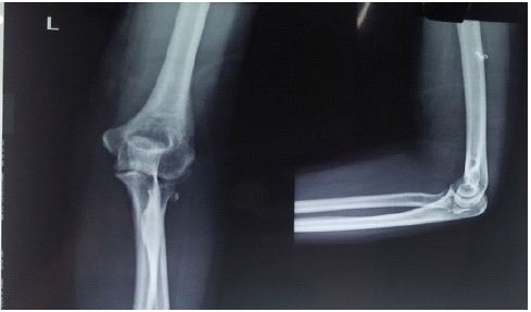
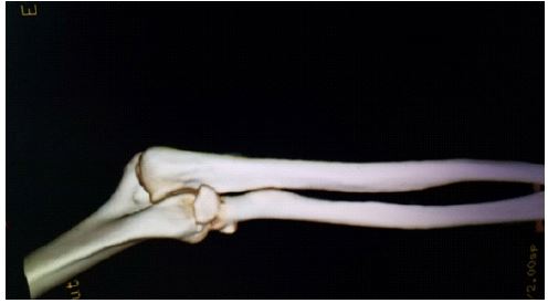
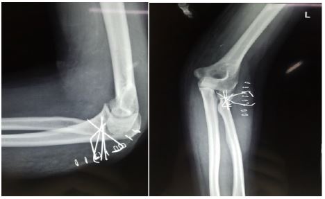
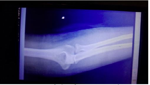
A 35 year old female was brought to our emergency department with complaints of pain and swelling over left elbow following history of fall from height on outstretched hand. Clinical and radiological (radiography (Figure 1) and 3D Computed Tomography (CT) scan) (Figure 2) evaluation demonstrated characteristics of Mason type –III fracture.
After pre-operative preparation, surgical intervention with open reduction and internal fixation was planned.
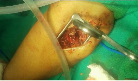
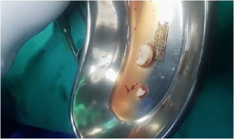
During the operation, as the commination made it difficult for classical ORIF, ex-situ radial head reconstruction and fixation was done with k-wire.
The patient recovered well and the k-wire was removed after 4 weeks postoperatively. Full range of motion (ROM) with normal function was achieved after physiotherapy.
Discussion
Various techniques for fixation of radial head fractures can be performed. Among them, ORIF is a very good option with satisfactory result but may be associated with complications which may require surgical interventions postoperatively. Radial head excision or radial head replacement with prosthesis is a much better option but is associated with reduction in joint mobility and rejection of the prosthesis.
Both these options can be difficult to perform in cases of severely comminuted fractures. In such cases, on-table reconstruction followed by K-wire fixation can be a very good option.
Kumar et al [9] and Businger et al [10] reviewed six cases (four cases of Mason type-III and four cases of type IV radial head fractures) managed with on-table reconstruction and fixation. They found that on-table reconstruction and fixation of comminuted radial head fractures provides a reasonably good result. Furthermore, there was no need for any other surgical intervention post-operatively regardless of the radiological findings. No patient complained of stiffness of the fingers or developed a complex regional pain syndrome, nor was there any objective evidence of instability of the elbow. Similar to these findings, our patient also had a very good range of motion on follow-up and had no complaints of joint pain and stiffness. So, on-table reconstruction with K-wire fixation can be considered as a very good option for radial head fractures where classical ORIF is not possible due to communition.
Conclusion
On-table reconstruction and K-wire fixation can be a very good option for Mason type-III radial head fracture where classical ORIF is not possible.
Declarations
Author contributions: DS conceptualized the study and was in charge of the case. DS and SD wrote the original manuscript and reviewed and edited the manuscript. PV and RPS reviewed and edited the manuscript.
Consent: Written informed consent was obtained from the patient.
Acknowledgements: None.
Conflict of interest: None.
References
- Tarallo L, Mugnai R, Rocchi M, Capra F, Catani F, et al. Mason type III radial head fractures treated by anatomic radial head arthroplasty: Is this a safe treatment option? Orthopaedics & traumatology, surgery & research: OTSR.. 2017; 103: 183-189.
- Jordan RW, Jones AD. Radial Head Fractures. The open orthopaedics journal. 2017; 11: 1405-1416.
- Mason ML. Some observations on fractures of the head of the radius with a review of one hundred cases. The British journal of surgery. 1954; 42: 123-132.
- Crönlein M, Zyskowski M, Beirer M, Imhoff FB, Pförringer D, et al. Using an anatomically preshaped low-profile locking plate system leads to reliable results in comminuted radial head fractures. Archives of orthopaedic and trauma surgery. 2017; 137: 789-795.
- Kaas L, van Riet RP, Vroemen JP, Eygendaal D. The epidemiology of radial head fractures. Journal of shoulder and elbow surgery. 2010; 19: 520-523.
- Bhandari M. Evidence-Based Orthopedics. Wiley-Blackwell. 2012.
- Mirzayan R, Itamura JM. Shoulder and Elbow Trauma. Thieme Medical Pub. 2004.
- Kachooei AR, Ring D. Evaluation of radiocapitellar arthritis in patients with a second radiograph at least 2 years after nonoperative treatment of an isolated radial head fracture. Arch Bone Jt Surg. 2017; 5(6).
- Jennings JD, Hahn A, Rehman S, Haydel C. Management of adult elbow fracture dislocations. Orthop Clin North Am. 2016; 47(1): 97–113.
- Ring D, Quintero J, Jupiter JB. Open reduction and internal fixation of fractures of the radial head. J Bone Joint Surg Am 2002; 84: 1811–1818.
- Kiran Kumar GN, Sharma G, Farooque K, Sharma V, Jain V, et al. On-table reconstruction and fixation of Mason type III radial head fractures. Chin J Traumatol. 2015; 18: 288-292.
- Businger A, Ruedi TP, Sommer C. On-table reconstruction of comminuted fractures of the radial head. Injury. 2010; 41: 583-588.