
Journal of Clinical Images and Medical Case Reports
ISSN 2766-7820
Clinical Image - Open Access, Volume 3
Disappearing left atrial thrombus with a hollow core
during anticoagulation therapy
Masahiro Kimura*; Keiko Maeda; Teruki Takeda; Hiroshi Mabuchi
Department of Cardiology, Koto Memorial Hospital, Higashiomi, Japan.
*Corresponding Author : Masahiro Kimura, MD, PhD
Department of Cardiology, Koto Memorial Hospital, 2-1Hiramatsu-cho, Higashiomi-shi, Shiga 527-0134, Japan.
Tel: (+81) 749-45-5000, Fax: (+81) 749-45-5001;
Email: mkimura@kuhp.kyoto-u.ac.jp
Received : Nov 01, 2022
Accepted : Nov 16, 2022
Published : Nov 23, 2022
Archived : www.jcimcr.org
Copyright : © Kimura M (2022).
Keywords: Atrial fibrillation; Atrial appendage; Thrombus.
Citation: Kimura M, Maeda K, Takeda T, Mabuchi H. Disappearing left atrial thrombus with a hollow core during anticoagulation therapy. J Clin Images Med Case Rep. 2022; 3(11): 2169.
Description
Left atrial thrombi with a hollow core are sometimes detected in patients with Atrial Fibrillation (AF) and Flutter (AFL) by Transesophageal Echocardiography (TEE). In previous studies, this finding at the initial assessment denotes relatively fresh and acute growing thrombus with inner liquefaction [1-3]. However, its significance during anticoagulation treatment is unknown.
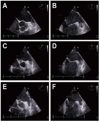
A 60-year-old man with AF and AFL admitted to our hospital because of heart failure. After 60 mg edoxaban once daily was administered for one week, a TEE was performed in advance of electrical cardioversion. Moderate spontaneous echo contrast was observed in left atrium and 11 mm × 12 mm isoechoic round thrombus in Left Atrial Appendage (LAA) was detected (Figure 1A and B), then cardioversion was cancelled. One week later, a repeated TEE was performed. The outer diameter was unchanged but echolucent area was appeared in the center of the thrombus (Figure 1C and D). Then anticoagulation therapy was continued. After 3 days, the thrombus was completely diminished assessed by TEE (Figure 1E and F). Brain Magnetic Resonance Imaging (MRI) and systemic contrast enhanced computed tomography revealed no thromboembolic event.
Recent studies have demonstrated the effectiveness of Direct Oral Anticoagulants (DOACs) for not only prevention but also treatment of LAA thrombus. In one prospective study, LAA thrombus resolution was seen in about 60% of patients treated with rivaroxaban for 6 weeks [4]. However, short-term sequential images of LAA thrombus dissolution during anticoagulation therapy are absent.
In our case, sequential TEE revealed that a hollow core of LAA thrombus was newly appeared during DOAC therapy just 3 days before complete dissolution. This echolucent change might reflect the process of clot lysis due to effective anticoagulational though thrombi usually shrink from the outside. In practice, when this clot appearance is observed for the first time during anticoagulation therapy, it is difficult to distinguish from fresh, unstable, and growing thrombus, therefore a careful follow-up is required. A TEE is a gold standard for detection and follow-up of LAA thrombus but some invasive, then other noninvasive modalities such as cardiac MRI are also considered for repeated examinations.
Conflict of interest: The authors declare that they have no conflict of interest.
References
- Singla V, Singh Y, Ravindranath SK, et,al. ‘Bird-beak sign’ of left atrial thrombus: A guide to management. BMJ case reports. 2013.
- Sinha A, Nanda NC, Khanna D, et al. Morphological assessment of left ventricular thrombus by live three-dimensional transthoracic echocardiography. Echocardiography. 2004; 21: 649-655.
- Dhawan S, Tak T. Left atrial mass: Thrombus mimicking myxoma. Echocardiography. 2004; 21: 621-623.
- Lip GY, Hammerstingl C, Marin F, et al. Left atrial thrombus resolution in atrial fibrillation or flutter: Results of a prospective study with rivaroxaban (X-TRA) and a retrospective observational registry providing baseline data (CLOT-AF). American heart journal. 2016; 178: 126-134.