
Journal of Clinical Images and Medical Case Reports
ISSN 2766-7820
Clinical Image - Open Access, Volume 3
Thoracic aortic pseudoaneurysm mimicking
recurrence of lung cancer
Masahiro Kimura1*; Fumihiro Miyashita2; Teruki Takeda1; Hiroshi Mabuchi1
1Department of Cardiology, Koto Memorial Hospital, Higashiomi, Japan.
2Department of Cardiovascular Surgery, Koto Memorial Hospital, Higashiomi, Japan.
*Corresponding Author : Masahiro Kimura, MD, PhD
Department of Cardiology, Koto Memorial Hospital, 2-1Hiramatsu-cho, Higashiomi-shi, Shiga 527-0134, Japan.
Tel: (+81) 749-45-5000, Fax: (+81) 749-45-5001;
Email: mkimura@kuhp.kyoto-u.ac.jp
Received : Nov 01, 2022
Accepted : Nov 17, 2022
Published : Nov 24, 2022
Archived : www.jcimcr.org
Copyright : © Kimura M (2022).
Keywords: Thoracic aorta; Pseudoaneurysm; Bias.
Citation: Kimura M, Miyashita F, Takeda T, Mabuchi H. Thoracic aortic pseudoaneurysm mimicking recurrence of lung cancer. J Clin Images Med Case Rep. 2022; 3(11): 2170.
Description
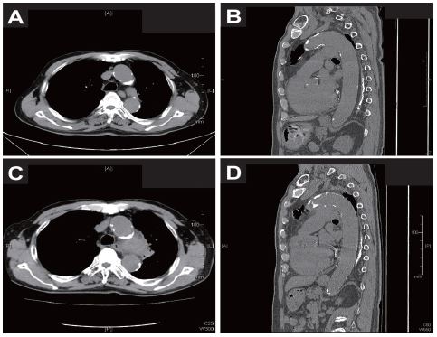
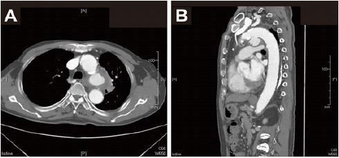
A man in his late 70’s visited to a hospital with one day history of fever and cough. Eight years before, he underwent a left upper lobectomy due to stage IB lung cancer. He had taken follow-up Computed Tomography (CT) exams annually and no abnormality was detected 9 months earlier (Figure 1A and B). Laboratory data showed elevated C-reactive protein, 9.7 mg/dL and D-dimer, 2.5 μg/mL with normal white blood cell counts. A chest CT on a visit showed a mediastinal mass near the resected site of left upper lobe (Figure 1C and D) with slight ground- glass opacity around it. Local recurrence of lung cancer with mild pneumonia was suspected, then an oral antibiotic was administered. His fever was alleviated and contrast-enhanced CT was performed one week later for detailed examination, which revealed a ruptured pseudoaneurysm of thoracic aorta (Figure 2A and B). Semi-urgent total arch replacement was completely achieved although it was tough due to severe adhesion of surrounding tissue. He was discharged without any complications on the 30th postoperative day.
Contained rupture of a thoracic aortic aneurysm is a rare condition but usually calls for an emergent repair because of high mortality rate. It is usually accompanied by a sudden-onset acute pain. We could not differentiate ruptured aneurysm from cancer recurrence by unenhanced CT images for the initial assessment. There are a few case reports of thoracic aortic aneurysm that was misrecognized as lung cancer [1,2] while lung cancer was also misrecognized as aortic aneurysm [3]. In our case, local adhesion due to previous surgery seemed to fortunately prevent complete rupture of thoracic aneurysm although postoperative state led to a cognitive bias for the misdiagnosis. Contrast-enhanced CT should be considered soon after the mass was detected in contact with aorta even if a typical chest or back pain is absent.
Conflict of interest: The authors declare that they have no conflict of interest.
References
- Che GW, Chen J, Liu LX, et al. Aortic arch aneurysm rupture into the lung misdiagnosed as lung carcinoma. Canadian journal of surgery. 2008; 51: E91-E92.
- Takahara Y, Nishiki K, Nakase K, et al. Ruptured pseudoaneurysm of the thoracic aorta mimicking lung cancer: A case report. Thoracic cancer. 2021; 12: 685-689.
- Lin F, Yang M, Guo C, et al. Lung cancer mimicking aortic dissecting aneurysm in a patient with situs inversus totalis. Thoracic cancer. 2016; 7: 254- 256.