
Journal of Clinical Images and Medical Case Reports
ISSN 2766-7820
Clinical Image - Open Access, Volume 3
Kaposi-Irgang disease: An entity not to ignore
Mouna Snoussi*; Fatma Mkaouar; Faten Frikha; Raida Ben Salah; Sameh Marzouk; Zouhir Bahloul
Internal Medicine, Hedi Chaker University Hospital, Sfax, Tunisia.
*Corresponding Author : Mouna Snoussi
Internal Medicine, Hedi Chaker University Hospital, Sfax, Tunisia.
Email: mounasnoussi23@gmail.com
Received : Sep 20, 2022
Accepted : Nov 28, 2022
Published : Dec 05, 2022
Archived : www.jcimcr.org
Copyright : © Snoussi M (2022).
Abstract
Kaposi-Irgang disease is a rare chronic from of cutaneous lupus. We describe a case of 28 years who was admitted for atrophic scars on both cheeks. The diagnosis of systemic lupus with chronic lupus profound was retained based on immunological and biopsy results. She was treated with hydroxychloroquine and referred to plastic surgery for lopfilling.
Citation: Snoussi M, Mkaouar F, Frikha F, Salah RB, Marzouk S, et al. Kaposi-Irgang disease: An entity not to ignore. J Clin Images Med Case Rep. 2022; 3(12): 2184.
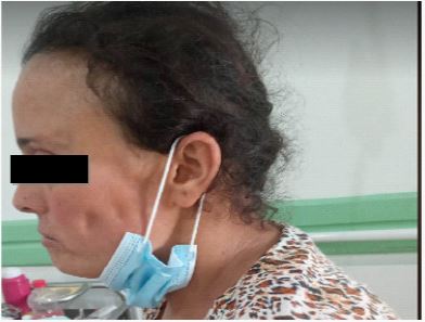
Clinical image description
A 28-year-old woman was admitted in internal medicine for arthralgia and skin lesions on the face. She reported a reccurent erythematous plaques in her both cheeks. She was initially managed by dermatologists and was given local corticosteroid therapy with no improvement. Physical examination found three atrophic scars on both cheeks of different sizes without inflammatory signs or induration (Figures 1 & 2). There were no other skin lesions. There were no arthritis or synovitis. Skin biopsy was performed and concluded to a superficial and deep dermal lymphocyte infiltration. Laboratory examination showed a mild lymphopenia at 1200 elts /mm³ in blood cell count and erythrocyte sedimentation rate was at 15 mmH [1]. The urine cast and the protinuria were negative and the creatinine was at 60 μmol/l. The anti-nuclear antibody were at 1/640 with anti Sm and anti nucleosome antibody positive. The diagnosis of systemic lupus was retained according to the ACR criteria revised in 1997. Cutaneous lesions corresponded to lupus profundus type or Kaposi-Irgang disease. The patient was treated with hydroxychloroquine at 400 mg/day for 3 months and then tapered to 200 mg/d. The patient was unsatisfied with the unsightly scars and was referred to plastic surgery department for a lipofilling.

References
- C. Francès et al. / Manifestations dermatologiques du lupus Dermatologic manifestations in lupus erythematosus. La Revue de médecine interne. 2008; 29: 701–709.
- Besma Ben Dhaou, Asma kefi, Zohra Aydi, Imen Rachdi, Houda Hammami, Fatma Daoud, Ehsen Ben Brahim, Sami Fenniche, Achraf Debbiche, Fatma Boussema. Lupus erythematosus panniculitis: A case report. Journal of Dermatology & Dermatologic Surgery. 2017; 21(2): 110-112.