
Journal of Clinical Images and Medical Case Reports
ISSN 2766-7820
Clinical Image - Open Access, Volume 3
Molar tooth sign: Joubert’s syndrome
Hajar Zebbakh*; Abir Lemrabet; Siham El Haddad; Latifa Chat; Nazik Allali
Department of Pediatric Radiology, Children’s Hospital, Mohamed V University, Rabat, Morocco.
*Corresponding Author : Hajar Zebbakh
Department of Pediatric Radiology, Children’s Hospital, Mohamed V University, Rabat, Morocco.
Tel: + 0616367908;
Email: drzebbakh93@gmail.com
Received : Nov 03, 2022
Accepted : Nov 30, 2022
Published : Dec 07, 2022
Archived : www.jcimcr.org
Copyright : © Zebbakh H (2022).
Citation: Zebbakh H, Lemrabe A, Haddad SE, Chat L, Allali N, et al. Molar tooth sign: Joubert’s syndrome. J Clin Images Med Case Rep. 2022; 3(12): 2188.
Introduction
A 5-year-old female child from a well-monitored, full-term pregnancy with no specific history of ataxia, hypotonia and global developmental delay.
Discussion
Joubert syndrome is an autosomal recessive malformation affecting the cerebellum and brainstem, with a pre- or neonatalon set and with out clear gender predominance. It is a molar malformation of the midbrain and hindbrain.
The clinical picture is polymorphic; during the first months of life infants may present hypotonia, respiratory rhythm disorders, abnormal eye movements, or facial dysmorphia and in early childhood may appear ataxia, delayed motor development, intellectual deficit. Other manifestations are rare: retinal dystrophy, nephronophthisia, polydactyly [1].
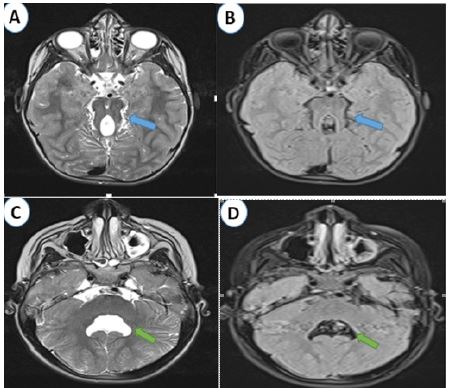
On MRI, the characteristic radiological sign is the molar sign [2] due to hypoplasia of the cerebellar vermis and malformations of the midbrain with a deep inter-peduncular fossa and thick and elongated upper cerebellar peduncles, as well as the bat-wing appearance of V4 (Figure 1).
Differential diagnoses arise when there is an abnormality of the vermis and V4. Joubert syndrome can be confused with Dandy walker malformation and Rhomboencephalosynapsis, but the molar tooth appearance of the midbrain is pathognomonic of Joubert syndrome.
No known treatment has been developed for this disorder, although some organizations are currently studying the disorder, its prevention and treatment.
References
- Romani M, Micalizzi A, Valente EM. Joubert syndrome: Congenital cerebellar ataxia with the molartooth. Lancet Neurol. 2013; 12: 894–905.
- Fluss J, Blaser S, Chitayat D, et al. Molartooth sign in fetal brain magnetic resonance imaging leading to the prenatal diagnosis of Joubert syndrome and related disorders. J Child Neurol. 2006; 21: 320–324.