
Journal of Clinical Images and Medical Case Reports
ISSN 2766-7820
Case Report - Open Access, Volume 3
Optical coherence tomography angiography (OCTA) findings in
a patient with acute traumatic choriocapillaropathy
Mohamadreza Ansari Astaneh; Maryam Hedayati; Mehrdad Motamed Shariati*
Eye Research Center, Mashhad University of Medical Sciences, Mashhad, Iran.
*Corresponding Author : Mehrdad M Shariati
Eye Research Center, Khatam Al-Anbia eye hospital, Gharani boulevard, Mashhad, Iran.
Tel: +98-937-738-8690;
Email: mehrdad_shariati2005@yahoo.com
Received : Nov 10, 2022
Accepted : Dec 01, 2022
Published : Dec 08, 2022
Archived : www.jcimcr.org
Copyright : © Shariati MM (2022).
Abstract
Acute traumatic maculopathy included a spectrum of disorders following blunt ocular trauma, such as traumatic pigment epitheliopathy, commotio retina, and choroidal rupture. Our patient was a 47-year-old male who came to the emergency department due to blurred vision in his left eye for the past two weeks following blunt ocular trauma. His best-corrected visual acuity was 10/10 in the unaffected right eye (OD) and 7/10 in the left eye. In fundus examination, we found decreased foveal reflex and gray-white spots in a ring-like pattern at the parafovea and an area of pigmentary change below the superior arcade near the disc. Optical Coherence Tomography (OCT) demonstrated disruption of the Ellipsoid zone, and Optical Coherence Tomography Angiography (OCTA) showed dark dot signals in choriocapillaris at the same site. As we showed in this article, traumatic choriocapillaropathy, which can be described as spots of flow disruption within choriocapillaris can occur without any injury to the retinal vasculature.
Keywords: Trauma; Maculopathy; Choriocapillaris; Optical coherence tomography angiography.
Citation: Astaneh MA, Hedayati M, Shariati MM. Optical coherence tomography angiography (OCTA) findings in a patient with acute traumatic choriocapillaropathy. J Clin Images Med Case Rep. 2022; 3(12): 2191.
Introduction
Blunt ocular trauma manifests in various ways in the posterior segment of the eye. It causes serious sequelae such as Commotio Retinae (CR), choroidal rupture, Traumatic Pigment Epitheliopathy (TPE), Purtscher’s retinopathy, macular holes, Vitreous hemorrhage, retinitis sclopetaria, retinal breaks, and retinal dialysis [1].
Spectral-Domain Optical Coherence Tomography (SD-OCT) imaging supports the idea that the primary site of injury is the photoreceptor and Retinal Pigment Epithelium (RPE) layers. Depending on the severity of the trauma, SD-OCT may reveal different optical densities of intraretinal spaces, which may range from mild lesions with transient hyper-reflectivity of the outer retina and disappearance of the thin hypo-reflective optical space; to more severe cases with areas of disruption in the Inner Segment/ Outer Segment (IS/OS) junction and hyper-reflectivity of the overlying retina, pigment disorders and retinal atrophy. Optical Coherence Tomography (OCT) is a powerful non-invasive tool for evaluating the retinal status [2].
OCT findings usually show disruption of the IS/OS junction (Ellipsoid zone) and corresponding hyperreflectivity, defects at the cone OS tips, or damage to the external limiting membrane [3].
Transient hyper-reflectivity of the outer retina in OCT imaging may associate with a good prognosis. More severe trauma may disrupt the IS/OS junction with hyper-reflectivity of the overlying retina, defects at the cone OS tips, damage to the external limiting membrane, pigment disturbance, and retinal atrophy, which corresponds to poor visual prognosis [2,3].
OCT Angiography (OCTA) is a non-invasive imaging technique that provides en face visualization of the retinal vascular networks by detecting the blood flow in different retinal layers and allowing for the investigation of the possible vascular involvement after ocular trauma, which may help detect the impairment of the retinal microvasculature and its progressive changes over time even in the absence of compromised visual acuity [4].
In this report, we will describe a macular OCTA image of a subject with acute traumatic maculopathy following blunt ocular trauma.
Case report
A 47-year-old male was referred to the emergency department due to blurred vision in his left eye for the past two weeks following blunt ocular trauma with a wooden rod. The subject underwent the following ophthalmic evaluations: Best-Corrected Visual Acuity (BCVA) measurement with thumbing E chart, slit-lamp biomicroscopy, Goldmann applanation tonometry, complete fundus examination ( using a +90D condensing lens), macular SD-OCT (Spectoralis®Tracking Laser Tomography (Heidelberg Engineering)) and OCTA (AngioVueRTVue XR Avanti, Optovue, Fremont, CA, USA, software version: 2018,0,0,18) with 6 x 6 mm scan size and macular fundus autofluorescence at the Khatam Anbia Eye Hospital. We discarded any images with a quality index below 6/10 and repeated the imaging. His best-corrected visual acuity was 10/10 in the unaffected right eye (OD) and 7/10 in the left eye. Relative Afferent Pupillary Defect (RAPD) was negative. Intraocular pressures were 10 and 8 mmHg in his right and left eyes, respectively. There were no limitations in eye movements. The right eye was normal in slit-lamp biomicroscopy and fund us examination. Slit-lamp examination of the left eye revealed no significant abnormality. In fund us examination, we found decreased foveal reflex and gray-white spots in a ring-like pattern at the parafovea and an area of pigmentary change below the superior arcade near the disc. There was no vitreous opacity, and the optic nerve head was normal.
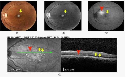
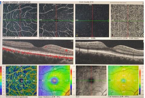
In the FAF image, we found hyperautofluorescent spots in a ring-like pattern and a patch of hyperautofluorescent lesion with a size of 1 disc diameter below the superior arcade compatible with fundus examination and red-free image. (Figure 1a,b,c). Optical Coherence Tomography (OCT) using Spectoralis®Tracking Laser Tomography (Heidelberg Engineering) demonstrated disruption of the Ellipsoid zone (Figure 1d), and Optical Coherence Tomography Angiography (OCTA) using the RTVue-XR Avanti system (Optovue Inc.) showed dark dot signals in the choriocapillaris (Figure 2) at the same location compared to the normal image of the other eye of the patient (Figure 3). Retinal vasculature seems to be intact following trauma.
After two months, ophthalmic evaluations showed no change in the imagings and visual acuity (Figure 4).

Discussion
Acute traumatic maculopathy included a spectrum of disorders following blunt ocular trauma, such as traumatic pigment epitheliopathy, commotio retina, choroidal rupture, and is a frequent cause of ophthalmic emergencies [2].
In this article, we introduced a 47-year-old male patient with decreased foveal reflex and gray-white spots in a ring-like pattern at the parafovea and an area of pigmentary change below the superior arcade near the disc two weeks after blunt ocular trauma. As we showed in the BAF and macular OCT images, there were hyperautofluorescent spots arranged in a ring-like pattern and a patch of hyperautofluorescent lesion below the superior arcade and ellipsoid zone disruption, which were the same size and at the same site as the lesions found in the fundus examination. We deduced two hypotheses for these hyperautofluorescent lesions: first, it could be due to loss of photoreceptor pigments that generally block the autofluorescent signals of the RPE cells [5], and second, it could happen because of metabolic stress in the RPE cells following shock wave injury. It seems that our first hypothesis is more compatible with this patient as we found areas of photoreceptor loss in the B-scan OCT. Daniel Lavinsky et al. in a case series in 2011, reported fundus blue autofluorescent and macular OCT findings in six eyes of six consecutive patients with blunt ocular trauma. They described a reduced FAF plaque with interposed increased FAF granular smaller lesions as traumatic pigment epitheliopathy [6]. Thuan et al. reported outer retinal thickening in the fovea with preservation of the inner retinal architecture and the foveal pit in the OCT images of 2 cases with acute traumatic maculopathy, which resolved within two weeks of the accidents [7].
In the OCTA image of our patient, we observed dark dot signals in the choriocapillaris consistent with the lesions, which we described previously, with intact retinal vasculature. It could be due to localized edema and vascular disruption or shadows of under-stress RPE cells. As we mentioned earlier, considering all the images, maybe the best explanation for dark dot signals in the choriocapillaris is localized edema and, or flow disruption. Optical Coherence Tomography Angiography is a non-invasive imaging technique for the evaluation of retinal and choroidal vasculature. It is safe, fast, and does not need any dye injection [8]. Today, the use of OCTA to evaluate retinal vasculature has increased. Yeter et al. in a study showed that the damage in the retinal microvasculature could be observed in traumatized eyes even with no findings in fundus examination [9]. There have been no reports about dark dot signals at the choriocapillaris in the literature.
Limitations
We had some limitations in this study. We evaluated the patient two weeks after the trauma due to the patient’s delay in coming. These findings do not belong to the acute phase of traumatic maculopathy, and somehow they are related to the complications of traumatic maculopathy.
Conclusion
As we showed in this article, traumatic choriocapillaropathy, which can be described as spots of flow disruption within choriocapillaris can occur without any injury to the retinal vasculature. Further studies are needed to understand the effects of blunt trauma on the choriocapillaris.
Declarations
Ethics approval and consent to participate: This study followed the tenets of the Declaration of Helsinki, and the Mashhad University of Medical Sciences ethical committee approved this study.
Consent for publication: We obtained informed consent from the patient to publish his examination results and images.
Conflict of interest: The authors declare any conflict of interests with the results of this paper.
Authors’ contributions: Conceptualization: Mehrdad Motamed Shariati, Mohammadreza Ansari Astaneh.
Patient examination and imaging: Maryam Hedayati, Mohammadreza Ansari Astaneh.
Writing the manuscript: Mehrdad Motamed Shariati, Maryam Hedayati.
Supervision: Mohammadreza Ansari Astaneh, Mehrdad Motamed Shariati.
References
- Guerra RLL, et al. Fundus autofluorescence in blunt ocular trauma. Arquivos brasileiros de oftalmologia. 2014; 77: 139-142.
- Mendes S, et al. Cutting edge of traumatic maculopathy with spectral-domain optical coherence tomography–A review. Medical Hypothesis, Discovery and Innovation in Ophthalmology. 2015; 4: 56.
- Mendes S, et al. Traumatic maculopathy 6 months after injury: A clinical case report. Case reports in ophthalmology. 2014; 5: 78-82.
- Montorio D, LD’Andrea, Cennamo G. Retinal Vascular Features in Ocular Blunt Trauma by Optical Coherence Tomography Angiography. Journal of Clinical Medicine. 2020; 9: 3329.
- Yung M, Klufas MA, Sarraf D. Clinical applications of fundus autofluorescence in retinal disease. International journal of retina and vitreous. 2016; 2: 1-25.
- Lavinsky D, et al. Fundus autofluorescence in patients with blunt ocular trauma. Acta ophthalmologica. 2011; 89: e89-e94.
- Pham TQ, et al. Optical coherence tomography findings of acute traumatic maculopathy following motor vehicle accident. American journal of ophthalmology. 2007; 143: 348-350.
- De Carlo TE, et al. A review of Optical Coherence Tomography Angiography (OCTA). International journal of retina and vitreous. 2015; 1: 5.
- Yeter DY, et al. Effect of blunt ocular trauma on retinal microvasculature: An optical coherence tomography angiography study. Photodiagnosis and Photodynamic Therapy. 2021; 33: 102147.