
Journal of Clinical Images and Medical Case Reports
ISSN 2766-7820
Case Report - Open Access, Volume 3
Aortic rupture during computed tomography
scanning: Even milliseconds matter
Açar Burak1*; Şimşek Uygur1; Karabacak Ali Rıza2; Umut Celikyurt1; Omay Oğuz3
1Department of Cardiology, Kocaeli University Medical Faculty, Kocaeli, Turkey.
2Department of Emergency Medicine, Kocaeli University Medical Faculty, Kocaeli, Turkey.
3Department of Cardiovascular Surgery, Kocaeli University Medical Faculty, Kocaeli, Turkey.
le*Corresponding Author : Acar Burak, MD
Department of Cardiology, Kocaeli University Medical Faculty, Umuttepe Yerleskesi, 41380, Kocaeli, Turkey.
Ph: +90 262 3038683, Fax: +90 262 3038003;
Email: burakacarmd@yahoo.com
Received : Nov 11, 2022
Accepted : Dec 02, 2022
Published : Dec 09, 2022
Archived : www.jcimcr.org
Copyright : © Burak A (2022).
Citation: Burak A, Uygur S, Rıza KA, Celikyurt U, Oğuz O, et al. Aortic rupture during computed tomography scanning: Even milliseconds matter. J Clin Images Med Case Rep. 2022; 3(12): 2193.
Background
A 52-year-old male patient was admitted to our emergency department with complaints of chest pain and back pain for one hour. The vital findings of the patient were normal except the blood pressure of between the arms. The right arm was of 170/110 mmHg and the left arm was of 140/90 mmHg. He was active smoker. The physical examination revealed diastolic murmur in the aortic area. Electrocardiography showed nonspecific findings. Transthoracic echocardiography showed aortic dissection and severe aortic insufficiency (Supplementary material). It was decided to perform Computed Tomography (CT) to see extension of dissection. At the beginning of the CT the patient was stable, and dissection was seen starting from aortic sinuses towards the iliac arteries (Figure 1A,1B,1C). During the procedure, only after 40 ms large pericardial effusion was detected just before venous phase starting which was compatible with aortic rupture and cardiac tamponade (Figure 1D). The patient was transferred for emergent surgery, however, he died during surgery.
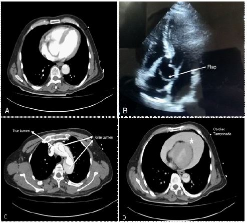
Figure 1: A. Computed tomography showing not apparent pericardial effusion B. Transthoracic echocardiography showing dissection flap C. Computed tomography showing dissection and false/true lumen at the level of aortic arch, D. Computed tomography revealing cardiac tamponade compression of heart chamber during CT scanning just before venous phase starting (after 40 ms).
Acute aortic syndromes are characterized bydissection, ulceration, or rupture of the thoracic aorta [1]. Intima tear of aorta cause penetration of blood into media. The gold standard method for diagnosis is CT [2,3]. Interestingly, aortic rupture occurred during CT scanning in this patient. We did not know whether it was related with CT scanning or not. Spontaneous rupture is fatal complication of aortic syndromes which could occur at any time during dissection.
Conflicts of interest: The authors have no conflicts of interest to declare.
References
- Morello F, Bima P, Castelli M, Nazerian P. Acute aortic syndromes: An internist’s guide to the galaxy. Eur J Intern Med. 2022; S0953-6205: 00355-00357.
- Ahn JM, Kim H, Kwon O, et al. Differential clinical features and long-term prognosis of acute aortic syndrome according to disease entity. European Heart Journal. 2019; 40: 2727–2736.
- Erbel R, Aboyans V, Boileau C, et al. 2014 ESC Guidelines on the diagnosis and treatment of aortic diseases: Document covering acute and chronic aortic diseases of the thoracic and abdominal aorta of the adult. The Task Force for the Diagnosis and Treatment of Aortic Diseases of the European Society of Cardiology (ESC). Eur Heart J. 2014; 35: 2873–2926.