
Journal of Clinical Images and Medical Case Reports
ISSN 2766-7820
Case Report - Open Access, Volume 3
Ortner syndrome secondary to left atrial dilatation
Mar Martínez-Ruiz-Coello1; Estefanía Hernández-García1,2; Estefanía Miranda1; Cristina García-García1; Ramón González-Herranz1,2; Guillermo Plaza1,2*
1Otolaryngology Department, Hospital Universitario de Fuenlabrada. Universidad Rey Juan Carlos, Madrid, Spain
2Otolaryngology Department, Hospital Universitario La Zarzuela, Madrid, Spain.
*Corresponding Author : Guillermo Plaza
Otolaryngology Department, Hospital Universitario de Fuenlabrada, Universidad Rey Juan Carlos, Madrid, Spain.
Otolaryngology Department, Hospital Universitario La Zarzuela, Madrid, Spain.
Email: gplaza.hflr@salud.madrid.org
Received : Nov 15, 2022
Accepted : Dec 05, 2022
Published : Dec 12, 2022
Archived : www.jcimcr.org
Copyright : © Plaza G (2022).
Abstract
Introduction: Ortner Syndrome (OS) is a disorder that presents with hoarseness due to compression of the Left Recurrent Laryngeal Nerve (LRLN) of vascular origin, such as cardiac pathology.
Methods: We present the case of a 65-year-old patient with a history of double mitral injury after percutaneous mitral valvuloplasty operated 30 years ago, without current follow-up by Cardiology, who attends an ENT clinic due to progressive hoarseness of months of evolution. The patient also reported frequent choking. Videoestroboscopic examination showed left vocal cord paralysis with a moderate closure defect. A CT scan of the neck and chest was requested, which revealed heart disease with mitral valve disease, showing a large increase in the size of the left atrium with calcifications in its posterior wall. She was referred to Cardiology Department and diagnosed with “paucisymptomatic severe mitral stenosis”. LRLN paralysis was treated with hyaluronic acid injection under local anesthesia in the office. Subjective improvement was confirmed by the patient using VHI-10 scale. The stroboscopic image also showed a compensation of the glottic closure.
Conclusion: OS presents with progressive hoarseness in patients with vascular problems that can compress the LRLN. Mitral valve disease is the most common cause, since it occurs with left atrial dilation that causes compression of the nerve. This syndrome can be accompanied by other symptoms derived from the compression of structures such as the esophagus. Local measures can be proposed such as hyaluronic acid injection on the left vocal fold, among other treatments.
Keywords: Nerve, paralysis, vocal cord, valve disease, dilatation, dysphonia, infiltration.
Abbreviations: OS: Ortner Syndrome; LRLN: left recurrent laryngeal nerve.
Citation: Martínez-Ruiz-Coello M, Hernández-García E, Miranda E, García-García C, González-Herranz R, Plaza G, et al. Ortner syndrome secondary to left atrial dilatation. J Clin Images Med Case Rep. 2022; 3(12): 2195.
Introduction
Ortner Syndrome (OS) is a disorder that causes hoarseness due to compression of the Left Recurrent Laryngeal Nerve (LRLN) of vascular origin, such as rheumatic atrial growth [1]. The LRLN can be compressed by cardiac and vascular structures along its long course. In addition, this nerve runs through the aortic arch posteriorly before ascending towards the tracheoesophageal groove, being more vulnerable to compression by other structures. OS has been described as a dilatation of the left atrium due to mitral stenosis that causes paralysis of the LRLN; however, other etiological causes have been studied, such as aortic aneurysms, pulmonary artery aneurysms, and aortic dissections. Although rare, dysphonia can occur as the only symptom in vascular pathologies such as those previously described [2].
This syndrome was first described in 1897 by Nobert Ortner, an Austrian doctor, in a patient with mitral valve stenosis and left atrial dilatation in context. In the last 100 years, different cardiopulmonary conditions associated with LRLN palsy have been described. For all these reasons, the syndrome can also be called cardiovocal syndrome [3].
Some rare causes of OS can be giant cell arteritis, penetrating aortic ulcer, patent ductus arteriosus, or pericardial effusion. It is more common in older men and, in specific cases; it can appear in children with congenital heart failure. It is difficult to find data on the incidence of this syndrome in the literature, being able to affirm that it is a rare entity with a low prevalence at present [4].
Case presentation
Case report
We present the case of a 65-year-old non-smoker and non-drinker woman, with the only remarkable history of a double mitral lesion post percutaneous mitral valvuloplasty operated 30 years ago, without current follow-up by Cardiology. This patient came to the ENT clinic due to progressive hoarseness that has lasted for months, which interferes with her quality of life. The patient also reported frequent choking on liquids and solids for weeks, accompanied by episodes of post-ingestion cough.
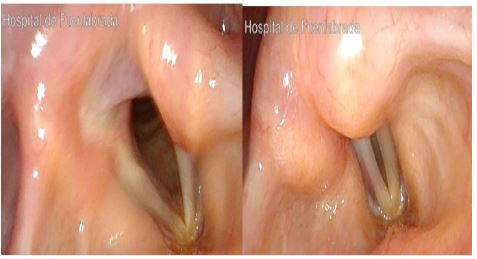
ENT examination by videostroboscopy showed paralysis of the left vocal cordin the paramedian position, with a moderate closure defect without other lesions or other findings (Figure 1 and 2, video 1). The glottic mucosal wave was present and laryngeal sensitivity was preserved. Cervical palpation did not reveal relevant findings.
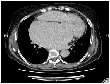
A Cervico-Thoracic CT scan revealed heart disease with mitral valve disease morphology manifested as a large increase in the size of the left atrium with calcifications in its posterior wall, as well as calcifications in the mitral valve area (Figure 3).
The patient had no dyspnea, orthopnea, or peripheral edema. She was referred to Cardiology consultations and diagnosed with “severe paucisymptomatic mitral stenosis without significant pulmonary hypertension” proposing follow-up by that same service.
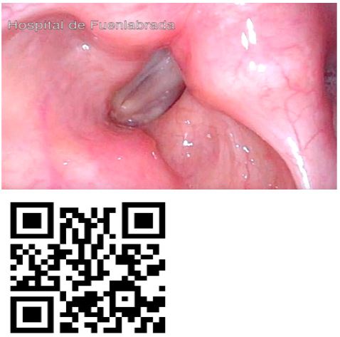
Left Recurrent Laryngeal Nerve (LRLN) paralysis was treated with hyaluronic acid injection on the affected vocal cord under in-office laryngeal infiltration of local anesthesia (Video 2). Subjective improvement was confirmed by the patient using the VHI-10 scale, whose results went from 23 pre-infiltration to 9 post-treatment. The stroboscopic image also confirmed an improvement in glottic closure and compensation (Figure 4 and video 3).


Discussion
When vocal cord paralysis is attributable to a cardiovascular process, it is called OS or cardiovocal syndrome. This was initially described by Nobert Ortner in 1897, defining left vocal fold paralysis caused by compression of the LRLN due to mitral stenosis. Since its initial description, OS has expanded to include other cardiovascular conditions, such as left atrial enlargement due to various cardiac or valvular abnormalities, pulmonary hypertension, aortic disorders, right subclavian artery aneurysm, and ductus arteriosus aneurysm [1-3]. This condition accounts for approximately 1.5% to 6.3% of all etiologies of unilateral vocal cord paralysis. To diagnose this entity, it is important to examine the entire course of the recurrent laryngeal nerves, including their course through the aortopulmonary window. The scan should extend inferiorly to include the pulmonary bifurcation [1-6].
An ENT examination is essential in patients with prolonged hoarseness. A persistent voice change of more than 2 weeks should be evaluated and studied, assessing the possible causes of hoarseness [4]. In addition to hoarseness, it should be noted that these patients usually suffer from dysphagia due to intrinsic compression of the esophagus. For this reason, it is important to perform imaging tests to rule out a compressive aetiology (neoplastic or vascular) in a patient with dysphagia [5].
Once vocal cord paralysis has been diagnosed as the cause of hoarseness by video fibroscopic examination, the etiology of the paralysis must be investigated. It is of the utmost importance to ensure the absence of suspicious lesions visible on ENT examination, including rigorous cervical palpation [4].
To rule out compressive causes of the paralysis, the study should be completed with cervico-thoracic imaging tests. The most widespread test today is CT, which is capable of showing us indirect signs that suggest vocal paralysis, such as medial displacement of the ipsilateral posterior margin of the vocal cords and ipsilateral laryngeal ventricular dilatation [7,8]. In addition, it allows us to evaluate the wide spectrum of cervical, cardiovascular or thoracic diseases that cause paralysis of the recurrent laryngeal nerve. The inclusion of the mediastinum up to the level of the aortopulmonary window (left side) or the brachiocephalic artery (right side) in CT studies performed for the etiological diagnosis of vocal fold paralysis is essential [8]. It is also possible to diagnose certain vascular compressive causes by means of a simple chest X-ray, with the advantages of similar sensitivity at a lower cost [9,10].
Regarding the cardiovascular causes that can compress the LRLN, there are several entities. The main cause defined was mitral valve disease with dilatation of the left atrium [1-6]. Subsequently, other pathologies have emerged that can cause this cardiovocal syndrome, such as: aortic aneurysms, pulmonary artery aneurysms and aortic dissection [10-13]. The incidence of cardiovocal syndrome varies depending on the underlying cause; in mitral stenosis it varies from 0.6% to 5%, while surgery for thoracic aortic aneurysm is associated with a relatively high incidence of about 32% [13]. In aortic dissection, chest pain is often referred to the back or abdominal area. The risk factors that predispose to aortic dissection are arterial hypertension, Marfan syndrome, aortic dilatation and coarctation of the aorta, among others [11].
Regarding treatment, in this case the patient was referred to the Cardiology Service with close follow-up without the need for surgical treatment at the present time. Regarding the treatment of LRLN paralysis, early injection of the paretic vocal cord with hyaluronic acid was decided, obtaining satisfactory results. Although there are controversies in the literature about the best time, material and technique to infiltrate the affected vocal cord, published data suggest that early intracordal injection may be useful in reducing pulmonary infections, length of hospital stay and the need for thyroplasty [14,15]. According to Wang et al. [15], the insufficiency of the glottic closure of infiltrated patients can improve, as well as the time of maximum phonation, increasing the performance of the vocal range. In addition, patients report a subjective improvement in their voice and quality of life. The duration of treatment effect is variable [15].
Conclusion
Ortner syndrome or cardiovocal syndrome is a rare entity that presents with paralysis of the LRLN due to extrinsic compression of cardiovascular aetiology. The most studied cause has been the pathology of the mitral valve, although in recent years other pathologies responsible for this syndrome have emerged, such as aortic and pulmonary aneurysms and aortic dissection. Its main symptom is hoarseness, although it can be accompanied by dysphagia. For its diagnosis, a correct anamnesis and a fibroscopic examination are essential to rule out malignant pharyngolaryngeal lesions. The study through imaging tests is essential to identify the compressive cause. Regarding the treatment of vocal paralysis, there are various options and techniques, with HA infiltration being a safe and effective alternative.
Declarations
The author(s) declared no potential conflicts of interest with respect to the research, authorship, and/or publication of this article.
References
- Kheok SW, Salkade PR, Bangaragiri A, Koh NSY, Chen RC, et al. Cardiovascular Hoarseness (Ortner’s Syndrome): A Pictorial Review. Curr Probl Diagn Radiol. 2021; 50: 749-754.
- Shrimanth YS, Barwad P, Maralakunte M, Sharma A, Sihag BK, et al. Ortner’s Syndrome Due to Giant Thoracic Aortic Aneurysm. J Invasive Cardiol. 2022; 34: E346.
- Subramaniam V, Herle A, Mohammed N, Thahir M. Ortner’s syndrome: case series and literature review. Braz J Otorhinolaryngol. 2011; 77: 559-562.
- Klee K, Eick C, Witlandt R, Gawaz M, Didczuneit-Sandhop B, et al. Unilateral recurrent nerve palsy and cardiovascular disease - Ortner’s syndrome. J Cardiol Cases. 2016; 15: 88-90.
- Ruiz-Serrato A, Pérez-Velasco MÁ, Guerrero-León Mde L, García-Ordóñez MÁ. Síndrome de Ortner y disfagia por aurícula izquierda gigante en el paciente anciano [Dysphagia and Ortnersyndrome due to a giantleftatrium in the elderly]. Rev Esp Geriatr Gerontol. 2015; 50: 204-205.
- Semionov A, Kosiuk J. Ortner syndrome secondary to aortic aneurysm. Radiol Case Rep. 2016; 12: 29-30.
- Bashir MH, Joyce C, Bolduan A, Sehgal V, Smith M, Charous SJ. Revisiting CT Signs of Unilateral Vocal Fold Paralysis: A Single, Blinded Study. AJNR Am J Neuroradiol. 2022; 43: 592-596.
- Paquette CM, Manos DC, Psooy BJ. Unilateral vocal cord paralysis: A review of CT findings, mediastinal causes, and the course of the recurrent laryngeal nerves. Radiographics. 2012; 32: 721-740.
- Leoce BM, Bernik JT, Voigt B, Dardik H, Bernik TR, et al. Ortner syndrome secondary to saccular thoracic aneurysm. J VascSurg Cases Innov Tech. 2021; 7: 371-373.
- Koh WJ, Azman M. Hoarseness in an older adult: Ortner syndrome. Malays Fam Physician. 2021; 16: 129-131.
- Hurtarte Sandoval AR, Carlos Zamora R, Gómez Carrasco JM, Jurado Ramos A. Ortner’s syndrome: a case report and review of the literature. BMJ Case Rep. 2014; 2014: bcr 2013202900.
- Srinivasan A, Agarwal R. Cardiovocal syndrome. Monaldi Arch Chest Dis. 2011; 75: 241-242.
- Plastiras SC, Pamboucas C, Zafiriou T, Lazaris N, Toumanidis S, et al. Ortner’s syndrome: A multifactorial cardiovocal syndrome. Clin Cardiol. 2010; 33: E99-100.
- Marques JAS, Marronnier A, Crampon F, Lagier A, Marie JP, et al. Early Management of Acute Unilateral Vocal Fold Paralysis: Update of the Literature. J Voice. 2021; 35: 924-926.
- Wang CC, Wu SH, Tu YK, Lin WJ, Liu SA, et al Hyaluronic Acid Injection Laryngoplasty for Unilateral Vocal Fold Paralysis-A Systematic Review and Meta-Analysis. Cells. 2020 5; 9: 2417.