Journal of Clinical Images and Medical Case Reports
ISSN 2766-7820
Clinical Image - Open Access, Volume 3
A case of Prune Belly syndrome associated to trisomy 21
*Corresponding Author : Narjiss Aji
Internal Medicine, Avicenne Hospital, Rabat,
Morocco.
Ph: +212610135516;
Email: narjissaji1998@outlook.fr
Received : Nov 14, 2022
Accepted : Dec 09, 2022
Published : Dec 16, 2022
Archived : www.jcimcr.org
Copyright : © Aji N (2022).
Citation: Aji N. A case of Prune Belly syndrome associated to trisomy 21. J Clin Images Med Case Rep. 2022; 3(12): 2202.
Clinical image description
We have actually in our unit a 18 months old boy admitted for another episode of pyelonephritis. He was born at full term by vaginal delivery with cephalic presentation and normal Apgar. His weight at birth was 3070 g.
His mother a 41-year-old Moroccan healthy women with 3 other normal children, had a poor antennal follow up in a tertiary care center. She does not report being related to the patient’s father.
The patient has presented a severe neonatal respiratory distress syndrome when he was 1 day year old and was hospitalized for 35 days in a private clinic. Two months later, the patient was hospitalized in our unit, for the first time, for a pyelo nephritis that was managed by anti biotherapy. The vital parameters were normal as follows: Temperature (T°) = 38.5°C; Breathing Rate (BR) = 58 Cycle per minutes; the Heart Rate (HR) = 148 Beats per minute; Blood Oxygen Saturation (SPO2) = 99%. For the anthropometric parameters: The height was 2DS below normal; the head circumference and the weight were 3DS below normal.
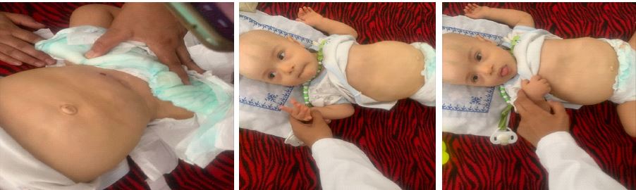
The conjunctivae and sclera were normal, a trisomic 21 facial was noted, the cardio-pulmonary exam was normal. The abdomen was distended (the ombilic perimeter was 50 cm). The abdominal wall seemed wrinkled. The intestines could be felt beneath the abdominal skin. The pulmonary examination is normal despite a pectus carinatum. The external genitalia were masculine, but the scrotum was empty, due to undescended tests. A balanic hypospadias was present. There was no sexual differentiation anomaly; the anus was present and permeable. The neurological exam found a hypotonic patient.
During his stay, the diagnosis of trisomy 21 have been confirmed by karyo type and a complete exploration looking for associated abnormalities have also been made and objective that the patient was presenting an atrioventricular canal defect and cerebral malformation. The kidney ultrasound report bilateral polycystic kidneys with a tortuous mega ureter.
Following the clinical triad: Cryptorchidism, abdominal distention and UTI malformations, the patient has been diagnosed with prune belly syndrome associated to trisomy 21. 4 months later, the patient has presented two repetitive pna, we would like to add that during his hospitalisation the patient has presented a pneumothorax. 5 months later the patient was operated for a urinary tract manipulation where a vesicostomy and ureterostomy have been made.