
Journal of Clinical Images and Medical Case Reports
ISSN 2766-7820
Case Series - Open Access, Volume 3
The interest of a hydrocolloid graft in the healing accompaniment of a subungual exostosis
Sokaina Chhiti*; Hanane Bayba; Fatima Zahrahashas; Zakia Douh; Meriam Soughi; Sara Elloudi; Fatima Zahra Mernissi
Dermatology Department, HASSAN II University Hospital, Fes, Morocco.
*Corresponding Author : Sokaina Chhiti
Resident, Dermatology Department, HASSAN II University Hospital, Fes, Morocco.
Ph: +21-269-830-8829;
Email: sokaina.chhiti@usmba.ac.ma
Received : Nov 21, 2022
Accepted : Dec 21, 2022
Published : Dec 28, 2022
Archived : www.jcimcr.org
Copyright : © Chhiti S (2022).
Abstract
Background: Subungual exostosis is a relatively uncommon, slow-growing benign bone tumor that most often affects the toes of young people and has a significant impact on quality of life. The treatment of which is essentially based on surgical excision with meticulous closure of the wound while avoiding the causes of nail dystrophy.
Material and methods: We report a new strategy for the management of the post-surgical wound at the nail level through an observational study in 4 young patients with a subungual exostosis treated by surgical excision followed by a hydrocolloid graft as post-surgical scar support with satisfactory results.
Results: 4 young girls whose average age was 19 years, consulted for a painful lesion at the nail level whose diagnosis of a subungual exostosis was confirmed clinically, radiologically and histologically and an excision surgery with placement of a hydrocolloid graft were successfully performed with a follow-up of one year
Conclusion: Postoperative complications of the wound in the context of subungual exostosis are significant and there is great variability in their appropriate management, hence the interest of codified post-surgical support based on hydrocolloid grafting.
Keywords: Subungual exostosis; Hydrocolloid graft; Wound; Management.
Citation: Chhiti S, Bayba H, Zahrahashas F, Douh Z, Soughi M, et al. The interest of a hydrocolloid graft in the healing accompaniment of a subungual exostosis. J Clin Images Med Case Rep. 2022; 3(12): 2218.
Introduction
Subungual exostosis is a rare benign bone tumor that mainly affects the toes of young people and can have a significant impact on quality of life [1]. The treatment consists of marginal excision and meticulous closure of the wound and well-conducted healing support to avoid postoperative complications.
Materials and methods
We report a new strategy for the management of the post-surgical wound at the nail level through an observational study conducted by our department and focused on 4 young patients with a subungual exostosis treated by surgical excision followed by a hydrocolloid graft as post-surgical scar support with satisfactory results over a period of 2 years.
Results
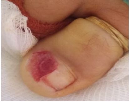
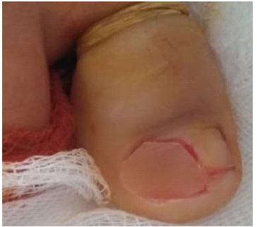
We collected 4 girls whose average age was 19 years old, consulted for a painful lesion under the nails when wearing or wearing tight shoes. Examination found a fixed, firm, tender 0.5 cm rounded nodule of normal skin color, keratotic surface below the nail plate (Figure 1). The X-ray was performed in all our patients, showed an opaque mass from the back of the distal phalanx (Figure 2). We performed, under local anesthesia, a marginal excision down to the normal bone without further disturbing the remaining nail bed (Figure 3). Histology was compatible with exostosis in all patients.


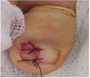
Our post-surgical healing support consisted of performing a hydrocolloid graft using two lateral stitches to fill the exposed nail bed, protect the distal phalanx and promote budding and healing (Figures 4,5,6). Hydrocolloid removal was performed after five days with application of fucidic acid, betadine tulles and a secondary dressing every day for 15 days, which was well tolerated by patients with rapid recovery and excellent postoperative aesthetic aspect (Figure 7).
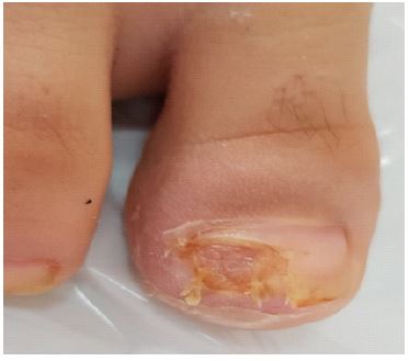
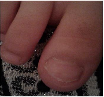
Our patients reported being asymptomatic after 6 weeks of follow-up and there was no sign of recurrence or onycholysis up to one year after resection with very satisfactory cosmetic results.
Discussion
Subungual exostosis is relatively rare and the initial diagnosis is often late or erroneous. Clinically, it presents as a firm, fixed nodule with a smooth, hyperkeratotic surface at the distal end of the last nail phalanx. Radiographs show a stalked radiopaque mass on the dorsomedial surface of the distal phalanx and the diagnosis is confirmed by history, physical examination, imaging, and histology. The mainstay of treatment is surgical excision [2]. However, postoperative complications can be a source of significant morbidity. In addition, several surgical techniques and postoperative wound closure strategies have been reported [3] to alleviate onychodystrophy and the recurrence associated with the treatment of this condition. Despite this, the rate of nail deformation remains high [4,5]. As Suga and Mukouda [6] reported in their review of postoperative nail deformity for lesions affecting the nail bed that were >5 mm, a direct approach was used and artificial skin was applied. Despite this, all these patients presented with onycholysis, which was not the case in our patients.
Although some attention has been given in the literature to strategies for optimizing surgical excision in the treatment of subungual exostosis, there are very few reports on optimizing postoperative wound management. Wounds can be treated by primary closure after resection, and some authors have described the possibility of secondary healing even after excision of part of the nail bed. The involvement of the nail bed and the size of the lesion play a role in the later development of onycholysis [3].
To address this issue, we report an effective technique based on hydrocolloid grafting in postoperative wound management in cases of subungual exostosis of the big toe. After marginal excision of the lesion, a sterile hydrocolloid dressing was applied to the wound directly against the phalanx bone using stitches so that the wound was covered in a sterile airtight environment. air and subjected to negative pressures to promote healing, wound contraction and minimize the risk of onycholysis. This is consistent with the principle of a Vacuum-Assisted Closing Device (VAC) [3].
Conclusion
We emphasize that the use of a hydrocolloid dressing in such settings may alleviate onycholysis, improve aesthetics, and offer the best chance of a rapid return to function.
Declarations
Consent: The examination of the patient was carried out in accordance with the principles of the Declaration of Helsinki.
Conflicts of interest: The authors declare no conflict of interest.
References
- Evison G, Prix CH. Exostose sous-unguéale. Br J Radio. 1966; 39: 451–455.
- Letts M, Davidson D, Nizalik E. Exostose sous-unguéale: diagnostic et traitement chez les enfants. Traumatisme. 1998; 44: 346–349.
- Da Cambra MP, Gupta SK, Ferri-de-Barros F. A novel management strategy for subungual exostosis. BMJ Case Rep. 2013; 2013: bcr2013200396.
- Multhopp-Stephens H, Walling AK. Exostose sous-unguéale: une technique simple d’exérèse. Pied Cheville Int. 1995; 16: 88–91.
- Lokiec F, Ezra E, KrasinE, et coll. Une technique chirurgicale simple et efficace pour l’exostose sous-unguéale. JPediatr Orthop. 2001; 21: 76–79.
- Suga H, Mukouda M. Exostose sous-unguéale. Ann Plast Surg. 2005; 55: 272–275.