
Journal of Clinical Images and Medical Case Reports
ISSN 2766-7820
Clinical Image - Open Access, Volume 3
Blue sea histiocytes detected in a patient with multiple myeloma
Bouatay Amina1*; Maatamri Wided2; Kortas Mondher2
1Hematology Laboratory, Sahloul University Hospital of Sousse, Tunisia.
2Hematology Laboratory, Farhat Hached University Hospital of Sousse, Tunisia.
*Corresponding Author : Bouatay Amina
Hematology Laboratory, Sahloul University Hospital of Sousse, 4000 Sousse, Tunisia.
Email: bouatayamina@yahoo.fr
ORCID ID: 0000-0003-0270-8013
Received : Nov 21, 2022
Accepted : Dec 22, 2022
Published : Dec 29, 2022
Archived : www.jcimcr.org
Copyright : © Amina B (2022).
Citation: Amina B, Wided M, Mondher K. Blue sea histiocytes detected in a patient with multiple myeloma. J Clin Images Med Case Rep. 2022; 3(12): 2220.
Clinical image description
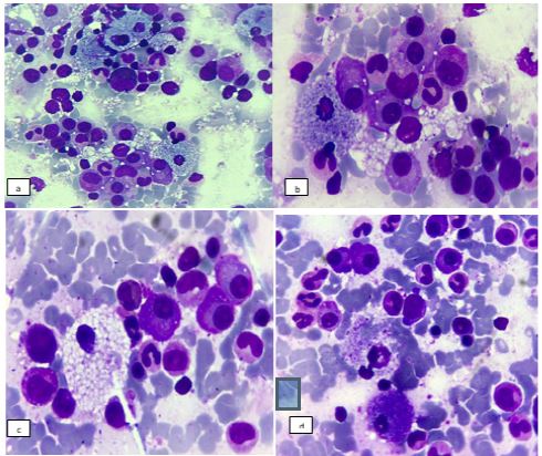
A 54-year-old man was admitted to the hematology department for exploration of splenomegaly. The blood count showed an isolated normochromic normocytic anemia (9.9 g/dL). Bone marrow aspiration showed a rich marrow with an estimated 35% plasma cell infiltration made up of dystrophic plasma cells (plasma cells with flamed cytoplasm, centered nuclei, multinuclearity), making the diagnosis of multiple myeloma. However, associated with this plasma cell infiltration, the marrow was infiltrated by abnormal histiocytes (Figure 1): Large foamy cells, filled with numerous small vacuoles and navy bluehistiocytes with dense and coarsely granular cytoplasm, raising the suspicion of a Niemann-Pick disease.
References
- Portier E, Talbot A, Nguyen Y, et al. Multiple myeloma occurring in a case of Niemann-Pick disease Type B: A pathophysiological link?. Br J Haematol. 2022; 197: e53-e55.