
Journal of Clinical Images and Medical Case Reports
ISSN 2766-7820
Case Report - Open Access, Volume 4
A very rare case: Early double-J stent fragmentation
*Corresponding Author : Emrah Yakut
Department of Urology, Ankara Memorial Hospital, Turkey.
Email: dremrahyakut@gmail.com
Received : Dec 05, 2022
Accepted : Dec 30, 2022
Published : Jan 06, 2023
Archived : www.jcimcr.org
Copyright : © Yakut E (2023).
Abstract
Background: The increasing use of a double-J (DJ) stent in urological diseases has also increased complications. In this report, we describe two cases presenting with early spontaneous DJ stent fragmentation.
Case presentation: The first patient was referred to our outpatient clinic due to pregnancy hydronephrosis. A DJ stent was placed in her right kidney. After 30 days, the stent was seen in the bladder in three fragments during cesarean section, and these fragments were removed. In the second patient, a DJ stent was placed in the right kidney following the detection of a stenosis in the right ureteropelvic junction. After 25 days, the patient visited our clinic again due to a stent fragment in urine. Fragmented DJ stent pieces were visualized on direct urinary system radiography.
Conclusion: Our cases once again demonstrated the need for a more careful and closer follow-up of all patients with stents.
Citation: Yakut E. A very rare case: Early double-J stent fragmentation. J Clin Images Med Case Rep. 2023; 4(1): 2231.
Introduction
The increasing use of Double-J (DJ) stents in urological diseases has also resulted in a significant increase in associated complications. Most of these complications are due to the prolonged stay of the DJ stent in the kidney. The spontaneous rupture of a DJ stent is a rare complication, but the removal of the remaining stent fragments from the kidney involves technical difficulties [1]. In this report, we describe two patients presenting with early spontaneous DJ stent fragmentation.
Case report
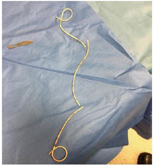
The first case was a 29-year-old female patient referred to our outpatient clinic due to pregnancy hydronephrosis. A DJ stent was placed in her right kidney. Thirty days later, she was taken to have a cesarean section, and the stent was planned to be removed in the same session. However, during this procedure, the DJ stent was seen in three fragments, which were then individually removed (Figure 1). No postoperative pathology was observed in the patient. The second patient was a 30-year-old woman presenting with flank pain. Examinations revealed a stenosis in the right ureteropelvic junction. She did not want to undergo surgery in the early period, and therefore a DJ stent was placed in her right kidney. After 25 days, she visited our clinic again after spotting a stent fragment in urine. Direct urinary system radiography revealed one DJ stent fragment in the renal pelvis and another in the bladder (Figure 2). Sponta Spontaneously fragmented DJ stent pieces were removed end oscopically. No postoperative pathology was observed in the patient. Both cases had a history of frequent urinary tract infections but no other commonality.

Discussion
Since the introduction of DJ stents in 1978, there have been many developments in stent composition and design. Stents are used to provide urinary drainage, allow stone passage, accelerate tissue healing, and prevent ureteral stenosis and urinary fistula formation. Stent use is simple, safe, and cost-effective method, but it does involve certain complications. Early stent complications include pain, bladder irritation, frequent urination, urinary incontinence, and fever, and late complications are petrification, migration, and fragmentation, which are more complicated and difficult to manage [2].
When a stent comes into contact with chemicals in urine, the tension, elasticity, and mobility of the stent decrease due to the degradation of stent polymers [3]. The optimal duration of stent stay is eight to 16 weeks. If a stent needs to be used for a longer period, stent replacement should be performed at intervals of eight to 12 weeks [4].
Generally, patients present with flank pain, hematuria, dysuria, and urinary excretion of stent fragments. In many cases, with or without urinary tract infection, leukocytes are found in complete urinalysis, which is considered to be due to stent depolymerization [5]. The stent material exposed to a specific environment within the body can be transformed from a bendable to brittle state [6].
Stent fragmentation is a relatively rare (0.3%) but major complication. In order to collect fragmented pieces endoscopically, the patient needs to be re-anesthetized, and this procedure presents with many surgical difficulties [7]. The transurethral route is usually sufficient to remove the stent. In some cases, extracorporeal shock wave lithotripsy, ureteroscopy, and percutaneous approaches may be necessary to fragment stones. Stent fragments in the middle ureter can be removed using a basket catheter under fluoroscopy [8].
The common feature of DJ stent fragmentation cases described in the literature is that almost all have had the stent for six months or longer. However, in the cases we presented, the stents were fragmented within one month. This early stent fragmentation can be associated with the patients’ history of frequent urinary tract infections, but there is not yet sufficient evidence. Nevertheless, these cases once again demonstrated the need for a more careful and closer follow-up of patients with stents.
References
- Zimskind PD, Fetter TR, Wilkerson JL. Clinical use of long-term indwelling silicone rubber ureteral splints inserted cystoscopically. The Journal of urology. 1967; 97: 840-844.Christodoulides AP, Karaolides T, Ibrahim ZM. Double J Stent Mislocation—Case Report. Open Access Library Journal. 2020; 7: 1-10.
- Christodoulides AP, Karaolides T, Ibrahim ZM. Double J Stent Mislocation—Case Report. Open Access Library Journal. 2020; 7: 1-10.
- Arshad M, Shah SS, Abbasi MH. Applications and complications of polyurethane stenting in urology. Journal of Ayub Medical College Abbottabad. 2006; 18: 69-72.
- Singh V, Gupta A. STENTURIA A RARE COMPLICATION OF INDWELLING URETERAL STENT (CASE REPORT). 2009.
- Ilker Y. Spontaneous fracture of indwelling ureteral stents in patients treated with extracorporeal shock wave lithotripsy: Two case reports. International urology and nephrology. 1996; 28: 15-19.
- Zisman A. Spontaneous ureteral stent fragmentation. The Journal of urology. 1995; 153: 718-721.
- El-Faqih S. Polyurethane internal ureteral stents in treatment of stone patients: morbidity related to in dwelling times. The Journal of urology. 1991; 146: 1487-1491.
- Kilciler M. Spontaneous ureteral stent fragmentation: A case report and review of the literature. The Kaohsiung journal of medical sciences. 2006; 22: 363-366