
Journal of Clinical Images and Medical Case Reports
ISSN 2766-7820
Short Report - Open Access, Volume 4
Radiolucency in conventional X-ray imaging leads potential diagnosis of hypoxia in human organ
Lopamudra Roy1; Amrita Banerjee2; Neha Bhattacharyya3,4; Susmita Mondal3; Ria Ghosh3; Manojit Das5,6; Radha Tamal Goswami7; Kallol Bhattacharya1; Asim Kumar Mallick8; Arpita Chattopadhyay7*; Samir Kumar Pal3*
1University of Calcutta, Department of Applied Optics and Photonics, JD-2, Sector-III, Salt Lake, Kolkata: 700 106, India.
2Department of Physics, Jadavpur University, Kolkata 700032, India.
3Department of Chemical and Biological Sciences, S.N.Bose National Centre for Basic Sciences, Block JD, Sector III, Salt Lake, Kolkata 700 106, India.
4Department of Radio Physics and Electronics, University of Calcutta, Kolkata: 700009, India.
5Department of Zoology, Uluberia College, University of Calcutta, Uluberia, Howrah-711315, India.
6Department of Zoology,Vidyasagar University,Rangamati, Midnapore-721102, India.
7Department of Basic Science and Humanities, Techno International New Town, Kolkata-700156, India.
8Department of Pediatric Medicine, Nil RatanSirkar Medical College and Hospital, 138, Acharya Jagadish Chandra Bose Rd, Sealdah, Raja Bazar, Kolkata, West Bengal 700014, India.
*Corresponding Author : Lena Marinova
Microbial Biotechnology Department, National Research Centre, Cairo, Egypt
Email: rad_marinova@abv.bg
Received : Dec 15, 2020
Accepted : Dec 30, 2020
Published : Dec 31, 2020
Archived : www.jcimcr.org
Copyright : © Marinova (2023).
Citation: Roy L, Banerjee A, Bhattacharyya N, Chattopadhyay A, Pal SK, et al. Radiolucency in conventional X-ray imaging leads potential diagnosis of hypoxia in human organ. J Clin Images Med Case Rep. 2023; 4(1): 2250.
Description
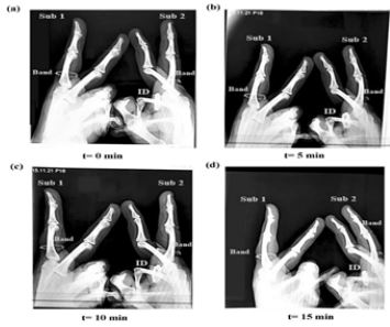
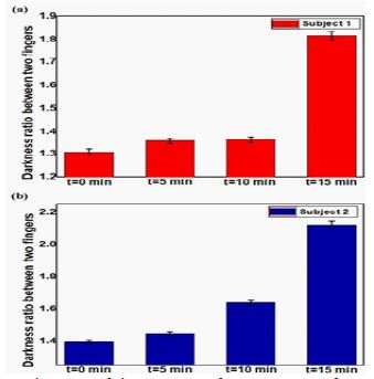
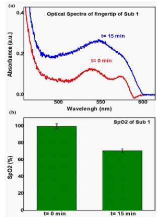
X-ray is one of the oldest, convenient, and cost effective imaging techniques commonly employed in the diagnosis of numerous clinical conditions. The differential X-ray absorption capacity, or contrast of the biological system is a determining factor in the biomedical X-ray picture interpretation. Due to the tendency of air and gas (such as oxygen, carbon-dioxide, nitrogen) to have low radiographic density, they tend to be radiolucent (i.e., not absorb X-rays) and hence, produce no shadow on X-ray film and appear black [1]. In the present work we have used radiolucent property of X-ray image of fingertips for the detection of controlled hypoxia generated due to blocking of blood circulation by using a rubber band. To our understanding, this work can serve as a template for development of the newer approaches to enhance the clarity of conventional X-ray radiography along with possibilities of detection of hypoxia in various organs of human subjects. Figure 1 depicts the X-ray images of fingertips of two subjects. For each subject index finger is blocked with rubber band in order to generate the partial hypoxia compared to middle finger at different time. The contrast of X-ray of the muscles (due to differential radiolucency) around fingertip is evident and their ratio are increasing with time (Figure 1, a-d). Figure 2 describes the ratio of the contrast of X-ray images around index and middle fingers of two subjects due to differential radiolucency indicating increasing hypoxia in the index finger as a result of partially blocked blood circulation by the rubber band. Optical absorption spectra using diffused reflection technique [2] of one of the subject’s index fingertip is shown in the upper panel of Figure 3. The absorbance around 560 nm and 576 nm indicate presence of deoxy and oxy-hemoglobin respectively in the target organ in different time. The estimated SpO2 values (details will be published elsewhere) are shown in the following panels of the Figure 3b. A clear correlation of the hypoxia detected through X-ray imaging (radiolucency) and optical technique is evident. Similar observation of manipulation of oxygen content in various target organ in pre-clinical mice model is reported earlier from this group [3]. To our understanding the technique would find its relevance for detection of hypoxia in various organs of human subjects in economically challenging countries. In particular, for the detection of severity due to diabetes-induced Peripheral Arterial Disorders (PAD) which is very common in present scenario would be very useful.
References
- Pfeiffer F, Bech M, Bunk O, Kraft P, Eikenberry EF, et al. Hard-X-ray dark-field imaging using a grating interferometer. Nature materials. 2008; 7: 134-137.
- N Bhattacharyya, S Singh, D Mukherjee, N Das, A Chatterjee, A Halder, et al. “Picosecond-resolved Fluorescence Resonance Energy Transfer (FRET) in Diffuse Reflectance spectroscopy explores biologically relevant hidden molecular contacts in a non-invasive way”, Phys. Chem. Chem. Phys. 2022; 24: 6176-6184.
- S Mondal, M Das, R Ghosh, S Singh, S Darbar, N Bhattacharyya, et al. Organ Specific therapeutic nanoparticles generates radiolucent reactive species for potential nanotheranostics using conventional X-Ray technique in mammals, Applied Nanoscience. 2022; 12: 3851–3858.