
Journal of Clinical Images and Medical Case Reports
ISSN 2766-7820
Short Report - Open Access, Volume 4
Labrune syndrome: A case Report
Zahra Ebadi; Elnaz Asadollahzade; Mohammad Ali Sahraian*
Multiple Sclerosis Research Center, Neuroscience Institute, Tehran University of Medical Sciences, Tehran, Iran.
*Corresponding Author : Mohammad A Sahraian
Sina MS Research Center, Sina Hospital, Tehran University of Medical Sciences, Hasan Abad Sq., Tehran, Iran.
Tel: 0098-21-66348571;
Email: sahraian1350@yahoo.com
Received : Jan 02, 2023
Accepted : Jan 23, 2023
Published : Jan 30, 2023
Archived : www.jcimcr.org
Copyright : © Sahraian MA (2023).
Abstract
Labrune disease or Leukoencephalopathy with brain calcifications and cysts (LCC) is a rare genetic disease. It distinguishes by a triad of leukoencephalopathy, calcifications, and intracranial cysts. Recently, a mutation in the SNORD118 gene has been identified as the genetic basis involved in the disease. A 34-year-old woman presented with progressive left hemiparesis. Neuroimaging reveals multiple parenchymal cysts, diffuse white matter hyperintensities, and scattered calcifications. These radiological findings after ruling out other differential diagnosis lead to the diagnosis of LCC.
Citation: Ebadi Z, Asadollahzade E, Sahraian MA. Labrune syndrome: A case report. J Clin Images Med Case Rep. 2023; 4(1): 2262.
Introduction
Labrune disease is a rare autosomal recessive genetic disorder recognized by Leukoencephalopathy with brain calcification and cysts (LCC). Mutation in SNORD118 leads to this disease [1,2]. The disease manifests in different ways such as seizure, cerebellar ataxia, and cognitive and brainstem impairment [1,3,4]. The age of onset is different and it can occur in childhood, young or late adulthood [5,6].
A set of three imaging findings including diffuse white matter hyperintense signal on T2-weighted imaging, cerebral calcifications, and brain cysts in brain Magnetic Resonance Imaging (MRI) help in diagnosis [1]. In this report, we present a young woman with leukoencephalopathy, supratentorial calcifications, and scatter parenchymal cysts in the brain MRI.
Case presentation
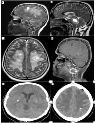
A 34-year-old woman, without a previous past medical history of any disease, presented with progressive left hemiparesis in February 2021. She denied any remarkable familial history or drug abuse. Physical exams were normal except for the decreased force of the left limbs. A gadolinium-enhanced brain MRI study showed multiple cystic lesions, most notably in the brain stem and subcortical white matter, and diffuse white matter hyperintensity on T2-weighted images (Figure 1). The wall of cysts and some subcortical lesions showed enhancement.
Various infection, neoplastic and inflammatory work-ups were done and all of them were negative. She was treated with steroids without any improvement. The patient underwent two brain biopsies, the first biopsy showed astrogliosis, Rosenthal fiber, wall thickening in vessels, and bright eosinophilic material. The other revealed fibrinoid material, infiltration of histiocytes, and degenerative change.
When she was referred to our clinic in June 2022, based on the cystic lesion on MRI and extensive involvement of white matter, a brain Computed Tomography (CT) scan was done. A brain CT scan reveals calcification in the deep cerebral nuclei. Due to the simultaneous presence of cystic lesions, calcification, and leukoencephalopathy, Labrune disease was diagnosed.
Discussion
More than 100 cases of LCC were reported since 1996 [7]. Labrune disease is cerebral microangiopathy [1,2]. Biallelic mutations in the SNORD118 gene are considered to cause LCC [2]. In neuroimaging, multiple cysts, diffuse extensive white matter involvement with sparing corpus callosum and U-fiber, and the presence of calcification lead to LCC diagnosis [8-10]. Calcification mostly is located in cerebral white matter or deep gray nuclei [11]. The cyst walls may be enhanced and also the mass effect is seen frequently [9]. The triad of extensive white matter T2 hypersignality, cerebral calcifications, and multiple cysts without the involvement of retinal or systemic presentation should propose the diagnosis of LCC [10].
Like our patient, in other Labrune-reported cases, sclerotic vessels with fibrinoid deposition, astrogliosis, Rosenthal fiber material, chronic microhemorrhage, and parenchymical calcification have been described [1,2,12,11,9,13,14,15].
Conclusion
In this article, we presented an adult-onset LCC. Clinical, radiological, and pathological findings are consistent with Labrune disease.
References
- Labrune P, Lacroix C, Goutiéres F, de Laveaucoupet J, Chevalier P, et al. Extensive brain calcifications, leukodystrophy, and formation of parenchymal cysts: a new progressive disorder due to diffuse cerebral microangiopathy. Neurology. 1996; 46: 1297-301.
- Jenkinson EM, Rodero MP, Kasher PR, Uggenti C, Oojageer A, et al., Mutations in SNORD118 cause the cerebral microangiopathy leukoencephalopathy with calcifications and cysts. Nat Genet. 2016; 48: 1185-1192.
- Corboy JR, Gault J, Kleinschmidt-DeMasters BK. An adult case of leukoencephalopathy with intracranial calcifications and cysts. Neurology. 2006; 67: 1890-1892.
- Chiang Y, Wang HJ, Chen CY. Adult-onset leukoencephalopathy, cerebral calcifications, and cysts: An 8-year neuroimaging follow-up of disease progression and histopathological correlation. J Clin Neurosci. 2019; 69: 276-279.
- Marelli C, Savoiardo M, Fini N, Bartolomei I, Marliani AF, et al. Late presentation of leucoencephalopathy with calcifications and cysts: report of two cases. J Neurol Neurosurg Psychiatry. 2008; 79: 1303-1304.
- Ummer K, Salam KA, Noone ML, Pradeep Kumar VG, Mampilly N, et al. Leukoencephalopathy with intracranial calcifications and cysts in an adult: Case report and review of literature. Ann Indian Acad Neurol. 2010; 13: 299-301.
- Osman O, Labrune P, Reiner P, Sarov M, Nasser G, et al. Leukoencephalopathy with calcifications and cysts (LCC): 5 cases and literature review. Rev Neurol (Paris). 2020; 176: 170-179.
- Sener U, Zorlu Y, Men S, Bayol U, Zanapalioglu U, et al. Leukoencephalopathy, cerebral calcifications, and cysts. AJNR Am J Neuroradiol. 2006; 27: 200-203.
- Ma Y, Zhang X, Cheng C, Xu Q, Di H, et al. Leukoencephalopathy with calcifications and cysts: A case report. Medicine (Baltimore). 2017; 96: e7597.
- Bonomo G, Monfrini E, Borellini L, Bonomo R, Arienti F, et al. Systemic involvement in adult-onset leukoencephalopathy with intracranial calcifications and cysts (Labrune syndrome) with a novel mutation of the SNORD118 gene. Eur J Neurol. 2020; 27: 2329-2332.
- Stephani C, Pfeifenbring S, Mohr A, Stadelmann C. Late-onset leukoencephalopathy with cerebral calcifications and cysts: case report and review of the literature. BMC Neurol. 2016; 16: 19.
- Kaffenberger T, Valko PO, von Meyenburg J, Baráth K, Hewer E, et al. A case of late onset leukoencephalopathy with cerebral calcifications and cysts in a 59-year-old woman. Eur J Neurol. 2009; 16: 278-281.
- Kleinschmidt-Demasters BK, Cummings TJ, Hulette CM, Morgenlander JC, Corboy JR et al. Adult cases of leukoencephalopathy, cerebral calcifications, and cysts: expanding the spectrum of the disorder. J Neuropathol Exp Neurol. 2009; 68: 432-439.
- Novo J, Lin D, Shanks M, Kocak M, Arvanitis L, et al. A 55-year-old female with leukoencephalopathy with cerebral calcifications and cysts: Case report and radiopathologic description. Pathol Res Pract. 2017; 213: 1440-1444.
- Tamura R, Ohira T, Emoto K, Fujiwara H, Horikoshi T, et al. Leukoencephalopathy, cerebral calcifications, and cysts: A clinical case involving a long-term follow-up and literature review. J Neurol Sci. 2017; 373: 60-65.