
Journal of Clinical Images and Medical Case Reports
ISSN 2766-7820
Case Report - Open Access, Volume 4
Purpura fulminans: A rare condition that surprisingly develops after endoscopic retrograde cholangiopancreatography
Serife Degirmencioglu Tosun1; Koray Koçhan1; Sercan Kiremitçi1; Ibrahim Hakkı Köker1; Hakan Senturk1,2*
1Bezmi Alem Vakıf University School of Medicine, İstanbul, Turkey.
2Department of Gastroenterology, Bezmi Alem VakıfUniversity, Topkapı mahallesi Fatih/ İstanbul, Turkey.
*Corresponding Author : Hakan Senturk
Department of Gastroenterology, Bezmi Alem VakıfUniversity, Topkapı Mahallesi Fatih/ İstanbul, Turkey.
Email: drhakansenturk@yahoo.com
Received : Jan 10, 2023
Accepted : Jan 24, 2023
Published : Jan 31, 2023
Archived : www.jcimcr.org
Copyright : © Senturk H (2023).
Abstract
Purpura Fulminans (PF) is a disease that occurs after septicemia. It has a rapid course. PF causes disseminated intravascular coagulation and severe organ failure. Extremity amputation and fatality rates are very high, even if it is diagnosed and treated promptly. This case report presented a 59-year-old man who developed purpura fulminans after Endoscopic Retrograde Cholangiopancreatography (ERCP).
Citation: Tosun SD, Koçhan K, Kiremitçi S, Köker IH, Şentürk H. Purpura Fulminans: A Rare Condition that Surprisingly Develops after Endoscopic Retrograde Cholangiopancreotography. J Clin Images Med Case Rep. 2023; 4(1): 2265.
Introduction
Purpura Fulminans (PF) is a rare disease with microvascular thrombosis and dermal hemorrhages affecting all age groups. High fever, tachycardia, and hypotension are the most common findings, and it progresses to peripheral circulatory collapse and shock in a short time. The mortality rate is very high. It often occurs during infection or within 2-4 week. PF is seen generally after Meningoccemia, infection with Haemophilus spp., and diseases such as chickenpox [1]. The lesions are sharply circumscribed, symmetrical and usually peripherally located. These lesions often turn into gangrene and require amputation. Sometimes, amputation can be prevented by extensive debridement and grafting [2].
Up to our knowledge this is the first article describing after Endoscopic Retrograde Cholangiopancreatography (ERCP). However, there are eight articles on the development of purpura fulminans due to E. coli. Most of these have been seen in the neonatal and childhood period. Two of them were seen in adulthood [3-8]. A specific strain of E. coli (Group A E. coli) was found in one case repot. Most PF cases developed after urinary tract infections and one case report was seen after heart transplantation [9]. We presented a case of fatal outcome due to PF after ERCP.
Case
ERCP was performed and a plastic stent had been placed for a 59-year-old male patient due to choledocholithiasis. The patient went to the same center for stent removal one year after the first procedure. The plastic stent was found migrated into the common bile duct. It could not be removed despite significant efforts, and he was referred to our center. The patient did not have any complaints at admission, and his physical examination and blood tests were within normal limits. His past history was, otherwise, negative. The migrated stent was removed with spyglass cholangioscope. He was well in the next morning, blood tests were normal and oral intake was started. The patient complained of sweating in the twentieth hour after the procedure, but no fever or hemodynamic changes were detected. However, it was observed that the previously normal CRP increased to 71 mg/L (reference range 0-5 mg/dl), and the procalcitonin level, which was not measured before, was 48 mg/L (baseline value < 0.5 mg/L). A blood sample was taken for culture. Parenteral fluid and intravenous antibiotics ceftriaxone 2 grams and metronidazole 3 x 500 mg) were started. Arterial blood pressure was measured as 85/60 mmHg, and pulse rate was 110 /min at the thirty-sixth hour. The lactate level in arterial blood was 13 mmol/L. Immediately after this, antibiotic treatment was changed to piperacillin-tazobactam (3 x 1.5 gr). In the meantime he was conscious, cooperative, and oriented. The abdominal examination revealed features a few ecchymoses around 2-3 cm in size on the periumblical region. Prolongation of peripheral arterial filling time was detected. He was taken to the intensive care unit to continiue his treatment the fortieth hour after procedure. Positive intropic treatment was started intravenously. Forty-eight hours after the procedure, rapid progressşon of ecchymosis on the fingertips, penis head, ear side, and nose was observed, and loss of consciousness developed. No pathology was detected to explain the situation on the computed tomography of the brain. Heparin infusion was started. White blood cell count was 54000 /uL (reference range 4000-10000 /uL), hemoglobin level was 9.96 g/dl (ref. ran.: 12-14 g/dl), platelet count was 57000 /uL (ref. ran.: 150000-450000 /uL) at the laboratory test. Vasculitic blood tests and coagulation parameters were analyzed due to the differential diagnosis of a rapidly progressive vasculitic picture, DIC (disseminated intravascular coagulation), sepsis, and TTP (thrombotic thrombocytopenic purpura). ANA, anti-ds DNA, protein C, protein S, anticardiolipin antibodies, anti-beta 2 glycoproteins 1 IgG, IgM, p-ANCA, and lupus anticoagulants were negative. Anti-thrombin 3 antigen was low, and antithrombin-3 activity was low. Escherichia coli growth in two blood cultures and only Acinetobacter baumani growth (sensitive to colistin) was detected in the tracheal aspirate. Colistin, meropenem, and vancomycin treatment were started. ERCP was performed two more times on the second and third days of his intensive care stay. Pus drainage was observed after biliary cannulation. The area was washed with antibiotics, and nasobiliary drainage was taken. On the fourth day of the intensive care follow-up, the patient was taken to Continuous Renal Replacement Therapy (CRRT) in the intensive care unit due to an anuric state. The ecchymotic areas of the patient turned into gangrene. Skin-punch biopsy revealed orthohyperkeratosis on the surface. Nearly complete necrosis in the epidermis, ischemic changes, and thrombosed vascular structures was seen in the upper dermis. Erythrocyte extravasation and solar degeneration were found. All these findings supported purpura fulminans. Plastic surgery consultation was requested when the demarcation line became evident in the ecchymosis areas of the patient. Plastic surgery recommended antibiotic therapy, keeping the limbs warm and wet dressing due to the development of granulation tissue in the demarcation line. They did not recommend escharotomy due to poor general condition. The patient was also consulted with hematology. His peripheral blood smear did not reveal any schistocyte or atypical cells. Hemolytic uremic syndrome or thrombotic thrombocytopenic purpura were not considered. The patient died on the 12th day of hospitalization despite the maximum antibiotic therapy, immunosuppressive therapy, therapy with protein C concentrates and Fresh Frozen Plasma (FFP), and inotropic support.
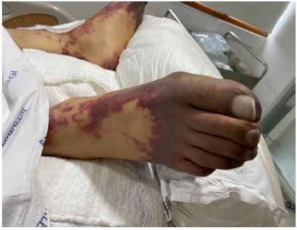
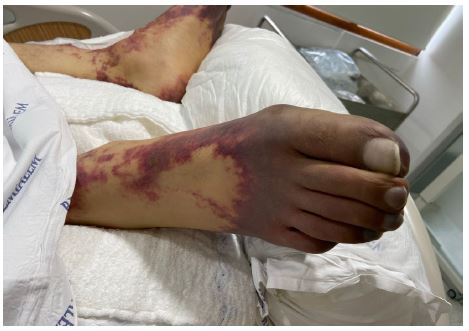
Table 1: The course of endoscopic treatments.
| Genetic factors -Protein C defficiency -Protein S defficiency -Factor V Leiden mutation -Abnormal complement level |
| Infectious diseases -Meningococ infection -Pneumococ infection -Haemophillus infection -Streptoccus species -Varicella infection -Capnocytophaga canimersus infection -Staphyloccus aureus infeciton |
| Autoimmun diseases -Autoimmune protein C and S defficiency |
| Cholestatic diseases -Cholangitis -Gall Blader infection |
| Drugs -Warfarin |
| Immune compromised diseases -HIV -Splenectomy before -Acquired abnormal complement level |
Discussion
Purpura Fulminans (PF) is a thrombotic type of Disseminated Intravascular Coagulation (DIC). Although PF is mainly seen in newborns, it may rarely be seen in adults as well. In most of the instances, it develops after an infection, mostly bacterial (either gram negative or positive), sometimes viral. Meningococcus is the most common cause of PF and its mortality rate is 15-25%. Haemophilus influenza, Capnocytophaga canimersus, streptococcus species, and staphylococcus aureus species have also been shown as other infectious causes of PF (Table 1). Dysfunction of endogenous anticoagulant pathways after infection is responsible for its pathophysiology. Inherited protein C (PC) and S (PS) deficiency have been associated with PF. PROC and PROS 1 genes were identified as responsible genes for PF as well. Decreased protein C, protein S, and antithrombin III levels due to cytokines activated by bacterial Autoantibodies against proteins C and S have been detected in patients with PF 7-10 days after infection. It has been observed that cross-reacting IgG-type antibodies increase the clearance of protein S. Protein C is a serine protease; it has anti-inflammatory and cytoprotective effects. Loss of endothelial thrombomodulin and decreased efficacy of activated protein C have been demonstrated in PF patients [10]. In a study by Lerolle et al., they compared the patients with Purpura fulminans and sepsis and those with sepsis and DIC. In the group with PF, bacteria in the vascular bed, bacterial endotoxins, changes in endothelial protein C receptor, change in CD 31 marker, and change in thrombomodulin were detected. Most PF patients are found to be positive for lupus anticoagulants. Asplenism, immunosuppressive diseases, heterozygous factor V Leiden mutation, alcohol use, and abnormal complement levels are other risk factors associated with PF [11].
In the early clinical presentation of PF patients, livedo reticularis appears on the extremities, ear, nose tips, back, and hips, following the latent period after infection. Afterward, areas of dermal vascular thrombosis and necrosis appear in the middle of these areas. If bleeding occurs in the necrosis areas, hemorrhagic bullae appear. Within 24-48 hours, full-thickness necrosis of the dermis settles. This feature differs from other diseases, such as ITP and TTP. The skin biopsy specimen can show thrombi within the dermal vascular structures histologically. End-organ damage is caused by vascular thrombosis. Later, hard eschars are formed. Bilateral, symmetrical involvement of the extremities is a typical finding. It is known that there is a very close relationship between the severity of skin lesions and protein C levels. Therefore, protein C concentrate or FFP should be started on patients without delay. Intravenous heparinization and, if necessary, antithrombin III can be used. Anti-coagulation and factor replacement therapies are at the most effective to prevent thrombosis-related end-organ damage. Thrombotic complications can occur long after the infection resolved. Mortality is very high. Most survivors have an amputation of the affected area [10-13].
In our case, the lesions were bilateral and symmetrical, with clear borders (Figures 1,2). The transformation of the post-infection lesions from livedo reticularis to necrosis developed within hours. Despite the administration of anticoagulants, anti-thrombotic therapy, protein C concentrate, and FFP in the early phase, necrosis and end-organ damage could not be prevented. Escharotomy, wound debridement, allografting, xenografting, or synthetic dressings can be used when the boundaries of necrosis become clear. It prevents fluid, electrolyte, protein loss, secondary bacterial infection, and increased catabolism can be prevented [10]. In our case, we tried to prevent this by us in wet dressing and keeping warm.
Warfarin-induced skin necroses, cryoglobulinemia, Anti-Phospholipid Syndrome (AFLS), Heparin-Induced Thrombocytopenia (HIT), and Henoch-Schonlein Purpura (HSP) should be considered in the differential diagnosis. All were discarded in our case.
Although E. coli is not frequently cited as the cause of PF, intestinal and extraintestinal E. coli related case with specific strain have been reported. In our case, Escherichia coli growth was detected in two sets of blood cultures. Broad-spectrum antibiotics were started. Two more biliary cannulations were performed after the first ERCP to ensure infection control, and pus drainage was provided. Although infection control was achieved, end-organ damage developed due to thrombotic complications, and the patient died.
Conclusion
Purpura fulminans is an acute, thrombotic, devastating emergency with high mortality. We are yet to know what exactly triggers purpura fulminans. It has been observed that some risk factors mentioned above lay the ground for this condition. Although early diagnosis and treatment reduces mortality, significant morbidity results. In summary, we described a patient developed Purpura fulminans after ERCP due to E. coli cholangitis who was lost despite timely exertion of all available management methods.
References
- Ergün SS, Atılganoğlu U, Kurşun I. Purpura fulminans: Olgu sunumu. Türkderm-Deri Hast Frengi Arşivi. 2002; 36: 292-294.
- Demir Z, Yüce S, Özdil K, Karamürsel S, Velidedeoğlu H, et al. Bir Purpura fulminans olgusu: Erken tanı ve tedavinin ekstremitelerin kurtulmasındaki önemi. Türkiye klinikleri journal of medical science. 2005; 25: 597-599.
- Lowry J, Noel E. A Rare Cause of a Rare Disorder: E. coli-Induced Purpura Fulminans Secondary to Urinary Tract Infection. Case Reports in Critical Care. 2022.
- Ahmed M, Samotowka M, Habis S, Mahmoud A, Saeed R. Escherichia coli bacteremia-induced purpura fulminans: a case report. Cureus. 2018; 10.
- Huemer GM, Bonatti H, Dunst KM. Purpura fulminans due to E. coli septicemia. Wiener klinische Wochenschrift. 2004; 116: 82-82.
- Adotama P, Savory S, Dominguez AR. Purpura fulminans in the setting of Escherichia coli septicemia. Cutis. 2015; 96: E3-E4.
- Hernandez MDMM, Carranza M, Patel B, Calvert J, Masri G. Purpura fulminans in a patient with septic shock due to Escherichia coli bacteremia with emphysematous pyelitis. Cureus. 2021; 13.
- Gürses N, Ozkan A. Neonatal and childhood purpura fulminans: review of seven cases. Cutis. 1988; 41: 361-363.
- Constant O, Vodovar D, Decousser JW, Mongardon N. Unusual purpura fulminans after heart transplantation. Intensive Care Medicine. 2019; 45: 887-888.
- Colling ME, Bendapudi PK. Purpura fulminans: mechanism and management of dysregulated hemostasis. Transfusion medicine reviews. 2018; 32: 69-76.
- Lerolle N, Carlotti A, Melican K, Aubey F, Pierrot M, et al. Assessment of the interplay between blood and skin vascular abnormalities in adult. 2013.
- Beechar VB, De La Flor C, Medford R J. Non-typeable Haemophilus influenzae and purpura fulminans. BMJ Case Reports CP. 2020; 13: e234880.
- Akoh CC, Singh G, Lederhandler M, Kim RH, Pomeranz MK. Purpura Fulminans Induced by Vibrio vulnificus. Cutis. 2021; 108: E7-E8.