
Journal of Clinical Images and Medical Case Reports
ISSN 2766-7820
Review Article - Open Access, Volume 4
Movement disorders associated with neurological tumors
Akram M Eraky1*; Nicole M Gregorich2
1Neurosurgery Department, Medical College of Wisconsin, Milwaukee, USA.
2School of Medicine and Public Health, University of Wisconsin-Madison, Madison, USA.
*Corresponding Author : Akram M. Eraky
Neurosurgery Department, Medical College of Wisconsin, Milwaukee, USA.
Email: aeraky@mcw.edu
Received : Dec 02, 2022
Accepted : Jan 30, 2023
Published : Feb 06, 2023
Archived : www.jcimcr.org
Copyright : © Eraky AM (2023).
Abstract
Neurological tumors, such as gliomas and meningiomas are rarely presented by movement disorders. Early detection of neurological tumors, both basal ganglia-arising tumors and basal ganglia-sparing ones, presenting with movement disorders is crucial to prevent any further deficits. In this review, we focus on neurological tumors-induced movement disorders, such as hemiballismus, hemichorea, myoclonus, Parkinsonism, and dystonia. These tumors affect dopaminergic pathways, basal ganglia pathways, or cerebellar nuclei by their mass effect or paraneoplastic impact. Our review highlights the importance of ruling out neurological tumors during the management of movement disorders.
Citation: Eraky AM, Gregorich NM. Movement disorders associated with neurological tumors. J Clin Images Med Case Rep. 2023; 4(2): 2272.
Introduction
Movement disorders are a rare manifestation of neurological tumors, such as gliomas and meningiomas through their effect on the dopaminergic pathways, basal ganglia pathways, or cerebellar nuclei by their mass effect or paraneoplastic impact [1,2]. Early detection of neurological tumors presenting with movement disorders is crucial to prevent any further deficits.
In this review, we classify neurological tumors causing movement disorders into basal ganglia-arising tumors, basal ganglia-sparing ones, pineal tumors compressing the midbrain, and tumors affecting cerebellar nuclei by its paraneoplastic effect. Moreover, we discuss the association between neurological tumors and movement disorders, such as hemiballismus, hemichorea, myoclonus, opsoclonus, Parkinsonism, and dystonia. Our review highlights the importance of ruling out neurological tumors during the management of movement disorders.
Neuroblastoma
Neuroblastoma (NB) is the most common malignant tumor in infants and arises from post-ganglionic sympathetic cells. Theoretically, it can derive from any sympathetic ganglion or adrenal medulla. It also occurs in association with neurofibromatosis type I [3,4]. NB has variable presentations that depend on its location, tumor-compressed structures, and aggressiveness varying from very aggressive to spontaneous regression [4].
NB arising from the adrenal medulla can be differentiated from Wilm’s tumor, another common malignant tumor in infants arising from the kidney, by NB’s tendency to cross the midline and displace the aorta [5]. Moreover, NB represents 50% of the causes of Opsoclonus myoclonus (OM) in infants which can be helpful in differentiating it from Wilm’s tumor. OM is an autoimmune condition affecting the cerebellar nuclei, especially the inhibitory Purkinje cells and excitatory granular neurons in the dorsal vermis through the paraneoplastic effect [6]. OM presents with myoclonus which is a quick sudden jerk of a group of muscles, opsoclonus which is nonrhythmic involuntary vertical and horizontal eye movements, and ataxia. Children with OM should immediately be investigated for neuroblastoma [6,7].
OM prognosis depends on its etiology; OM due to infection or idiopathy has a favorable prognosis. In contrast, cancer-induced OM has a bad prognosis and usually stays chronic [6]. OM treatment depends on eliminating the cause and prescribing symptomatic treatment, such as steroids, plasmapheresis, and immunosuppressive agents [6,7].
Pineal gland tumors
The pineal gland is a neuroendocrine gland secreting melatonin. It is surrounded superiorly by the corpus callosum splenium, laterally by the thalamus, and inferiorly by the superior colliculi of midbrain [8,9]. Pineal tumor manifestations are caused by mass effect and may vary according to their sizes, types, and compressed structures. Children are most commonly affected [8,9].
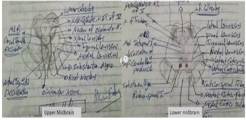
The most common pineal tumor is germinoma. Pineal germinoma can secrete hCG leading to precocious puberty in children. Also, large pineal glands can block cerebrospinal fluid through the aqueduct of Sylvius, causing obstructive hydrocephalus and increased intracranial pressure [8,9]. Typical physical examination shows a limitation of upward gaze and light-near dissociation which means the patient’s pupils are reactive to accommodation but not to light. This abnormal physical examination is known as parinaud syndrome or dorsal midbrain syndrome. Parinaud syndrome results from pressure on the pretectal region of the midbrain near the superior colliculus, Edinger-Westphal nuclei, and the oculomotor nerve (Figure 1) [8-11].
Basal ganglia and thalamus-arising tumors
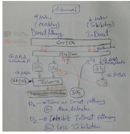
Basal ganglia includes caudate nucleus, putamen, globus pallidus, and subthalamic nucleus. These structures are connected together and to the substantial nigra. These networks form direct and indirect pathways organizing muscles’ movements (Figure 2) [12]. Any lesion affecting these basal ganglia will affect the presynaptic dopaminergic neurons or postsynaptic neurons and may result in many movement disorders, such as Parkinson’s disease, Huntington’s disease, hemiballismus, and dystonia [12].
Focal lesions affecting the subthalamic nucleus can cause hemichorea- hemiballismus presenting with unilateral dancing high-amplitude movement of an entire limb. Non-ketotic hyperglycemia, infarction and hemorrhage represents the most common causes [13-15]. Brain tumors, such as metastases and gliomas are rare causes of the hemiballismus [14,15]. Of interest, Donmez et al. reported a case of putaminal cavernous angioma causing hemichorea [16].
Tumors arising from caudate nucleus, putamen, or thalamus can cause hemidystonia presenting with unilateral involuntary muscle contraction including the contralateral arm, leg, and face [17]. Additionally, they can cause parkinsonism which manifests with bradykinesia, rigidity, and tremors by compressing the terminal ends of the substantia nigra neurons [1].
Basal ganglia-sparing supratentorial tumors
In a prospective study of 907 patients with supratentorial tumors, Krauss et al. found that parkinsonism incidence in those patients is about 3% and the most common tumor causing parkinsonism is meningioma [2]. Moreover, cerebellopontine angle tumors are reported as a rare cause of hemifacial spasm [18]. Of interest, many reports showed that frontal lobe tumors can cause parkinsonism, hemichorea, or dystonia [2,19,20]. This can be due to the mass effect of these tumors especially the large ones and their compression effect on the midbrain or basal ganglia [1].
Conclusion
In this review, we highlight the association between movement disorders and neurological tumors. Clinicians are encouraged to order neuroimaging during the management of any movement disorder to rule out neoplastic causes. Larger multicentric studies are encouraged to evaluate the importance of ruling out brain tumors in patients with movement disorders.
Declarations
Acknowledgment: Not applicable
Conflict of interest : The authors have no relevant financial or non-financial interests to disclose.
Funding: No funding was received for conducting this study.
Ethics approval and informed consent: Not applicable
References
- Marsili L, Vogrig A, Colosimo C: Movement Disorders in Oncology: From Clinical Features to Biomarkers. Biomedicines. 2021, 10: 26. 10.3390/biomedicines10010026
- Krauss JK, Paduch T, Mundinger F, Seeger W: Parkinsonism and rest tremor secondary to supratentorial tumours sparing the basal ganglia. Acta neurochir. 1995; 133: 22-9. 10.1007/BF01404943
- Shah S, Ravindranath Y: Neuroblastoma. Indian J Pediatr. 1998; 65: 691-705. 10.1007/BF02731044
- Davidoff AM. Neuroblastoma. Seminars in Pediatric Surgery. 2012; 21: 2-14. 10.1053/j.sempedsurg.2011.10.009
- Davenport KP, Blanco FC, Sandler AD: Pediatric malignancies: neuroblastoma, Wilm’s tumor, hepatoblastoma, rhabdomyosarcoma, and sacroccygeal teratoma. Surg Clin North Am. 2012; 92: 745-67. 10.1016/j.suc.2012.03.004
- Scarff JR, Iftikhar B, Tatugade A, Choi J, Lippmann S: Opsoclonus myoclonus. Innov Clin Neurosci. 2011; 8: 29-31.
- Rossor T, Yeh EA, Khakoo Y, et al.: Diagnosis and Management of Opsoclonus-Myoclonus-Ataxia Syndrome in Children: An International Perspective. Neurol Neuroimmunol Neuroinflamm. 2022; 9: 1153. 10.1212/NXI.0000000000001153
- Dahiya S, Perry A: Pineal Tumors: Advances in Anatomic Pathology. 2010; 17: 419-27. 10.1097/PAP.0b013e3181f895a4
- Favero G, Bonomini F, Rezzani R: Pineal Gland Tumors: A Review. Cancers. 2021; 13: 1547. 10.3390/cancers13071547
- Hoehn ME, Calderwood J, O’Donnell T, Armstrong GT, Gajjar A: Children with dorsal midbrain syndrome as a result of pineal tumors. Journal of American Association for Pediatric Ophthalmology and Strabismus. 2017; 21: 34-8. 10.1016/j.jaapos.2016.09.024
- Sciacca S, Lynch J, Davagnanam I, Barker R: Midbrain: Pons, and Medulla: Anatomy and Syndromes. RadioGraphics. 2019; 39: 1110-25. 10.1148/rg.2019180126
- Albin RL, Young AB, Penney JB: The functional anatomy of basal ganglia disorders. Trends in Neurosciences. 1989; 12: 366-75. 10.1016/0166-2236(89)90074-X
- Marsili L, Gallerini S, Bartalucci M, Marotti C, Marconi R: Paroxysmal painful spasms associated with central pontine myelinolisis in the context of nonketotic hyperglycemia. Journal of the Neurological Sciences. 2018; 388:37-9. 10.1016/j.jns.2018.03.005
- Glass JP, Jankovic J, Borit A: Hemiballism and metastatic brain tumor. Neurology. 1984; 34: 204-204. 10.1212/WNL.34.2.204
- Patankar AP: Hemi-chorea: an unusual presentation of brainstem glioma. British Journal of Neurosurgery. 2013; 27: 256-8. 10.3109/02688697.2012.741738
- Donmez B, Çakmur R, Uysal U, Men S: Putaminal cavernous angioma presenting with hemichorea: Clinical/Scientific Notes. Mov Disord. 2004; 19: 1379-80. 10.1002/mds.20207
- Chuang C: The natural history and treatment of acquired hemidystonia: report of 33 cases and review of the literature. Journal of Neurology, Neurosurgery & Psychiatry. 2002, 72:59-67. 10.1136/jnnp.72.1.59
- Colosimo C, Bologna M: Lamberti S, et al.: A Comparative Study of Primary and Secondary Hemifacial Spasm. Arch Neurol. 2006; 63: 441. 10.1001/archneur.63.3.441
- Saleh C, Akhalbedashvili N, Hund-Georgiadis M: Brain Tumor Presenting with Parkinsonism. Case Rep Neurol. 2021; 13: 595-7. 10.1159/000518198
- Rana A, Yousuf M, Hashmi M, Kachhvi Z: Hemichorea and dystonia due to frontal lobe meningioma. J Neurosci Rural Pract. 2014; 5: 290. 10.4103/0976-3147.133611