Journal of Clinical Images and Medical Case Reports
ISSN 2766-7820
Clinical Image - Open Access, Volume 4
The hole that saves the eye with no delay
Taoufik Abdellaoui1*; Yassine Mouzari2; Abdelbarre Oubaaz2
1Ophthalmology Department, Mohamed V Military Teaching Hospital of Rabat Sidi Mohamed Ben Abdellah University, Morocco.
2Ophthalmology Department, Mohamed V Military Teaching Hospital of Rabat Mohamed V university, Rabat, Morocco.
*Corresponding Author : Taoufik Abdellaoui
Ophthalmology Department, Mohamed V Military Teaching Hospital of Rabat Sidi Mohamed Ben Abdellah University, Fes, Morocco.
Email: taoufik.abdellaoui@hotmail.com
Received : Jan 17, 2023
Accepted : Feb 02, 2023
Published : Feb 09, 2023
Archived : www.jcimcr.org
Copyright : © Abdellaoui T (2023).
Citation: Abdellaoui T, Mouzari Y, Oubaaz A. The hole that saves the eye with no delay. J Clin Images Med Case Rep. 2023; 4(2): 2279.
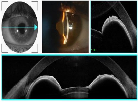
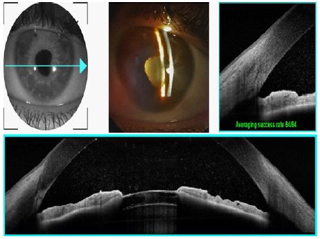
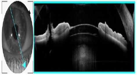
Clinical image description
A 28-year-old patient was followed up for unilateral anterior synechial uveitis of the right eye, with an inconclusive etiological work-up. Multiple relapses resulted in posterior synechiae formation around the pupil with a tomato iris appearance (Figure 1). The intraocular pressure was 45 mmHg. After preparation of the patient with intravenous mannitol infusion and hypotonic and corticosteroid eye drops, Peripheral Iridotomy (PI) with Yag laser was performed. Aqueous humor flow through the iridotomy orifice resulted in an immediate change in the curved iris configuration and separation of the iridotrabecular contact at the iridocorneal angle (Figures 2 and 3).