Journal of Clinical Images and Medical Case Reports
ISSN 2766-7820
Clinical Image - Open Access, Volume 4
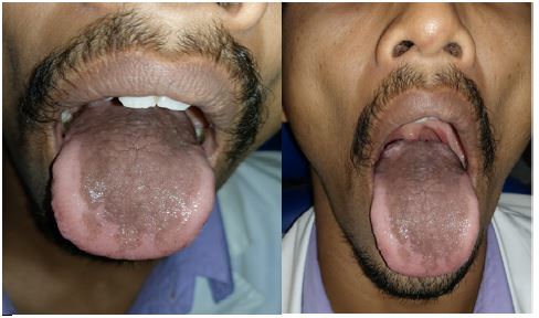
Physiologic pigmentation of dorsal surface of tongue
Prasanna R Sonar1*; Aarati Panchbhai2
1Resident, Sharad Pawar Dental College, DMIMS (DU), India.
2Professor, Sharad Pawar Dental College, DMIMS (DU), India.
*Corresponding Author : Prasanna Ravindra Sonar
Resident, Sharad Pawar Dental College, DMIMS (DU), India.
Ph: 8600719946;
Email: jaysonar1234@gmail.com
Received : Jan 03, 2023
Accepted : Feb 06, 2023
Published : Feb 13, 2023
Archived : www.jcimcr.org
Copyright : © Sonar PR (2023).
Citation: Sonar PR, Panchbhai A. Physiologic pigmentation of dorsal surface of tongue. J Clin Images Med Case Rep. 2023; 4(2): 2283.
Description
Pigmentation of oral mucosa is caused by pigments leading to change in colour of tissues. It can be endogenous or exogenous. Melanin, haemoglobin, hemosiderin, and beta-carotene are responsible for endogenous pigmentations. The majority of brownish pigmentation is physiological, however occasionally it might be a sign of serious disorders. Clinical manifestations of physiological pigmentation of the oral mucosa include multifocal or diffuse melanin pigmentation, which varies in prevalence across different racial and ethnic groups. The patient may not become aware of the patient’s physiological colouring until much later in life. Brown in colour, ranging from light to dark. Early in intrauterine development, melanoblasts, which are progenitors of melanocytes, go from the neural crest to the epidermis and hair follicles before differentiating into dendritic cells. After roughly 10 weeks of gestation, melanocytes first emerge in the head and neck region of the body.
Intra-oral examination of this patient shows diffuse, well demarcated, smooth, brownish pigmented lesion, present on dorsum surface of the tongue which is non tender and non-scrapable.