Journal of Clinical Images and Medical Case Reports
ISSN 2766-7820
Clinical Image - Open Access, Volume 4
Spontaneous pneumothorax in a severe case of emphysema
Mariana Fidalgo*; Inês Soares
Internal Medicine Resident, Centro Hospitalar Vila Nova Gaia / Espinho – Portugal.
*Corresponding Author : Mariana Fidalgo
Internal Medicine Resident, Centro Hospitalar Vila Nova Gaia / Espinho – Portugal.
Email: marianafidalgo@gmail.com
Received : Jan 25, 2023
Accepted : Feb 09, 2023
Published : Feb 16, 2023
Archived : www.jcimcr.org
Copyright : © Fidalgo M (2023).
Abstract
Pneumothorax is defined as the accumulation of air in the pleural cavity and typically presents as sudden chest pain and dyspnea. It is a complication of pulmonary emphysema and the likelihood of occurrence correlates with the severity of the underlying disease. It may present atypically, leading to delays in diagnosis and treatment.
Keywords: Pneumothorax; Emphysema; Smoking.
Citation: Fidalgo M, Soares I. Spontaneous pneumothorax in a severe case of emphysema. J Clin Images Med Case Rep. 2023; 4(2): 2290.
Case presentation
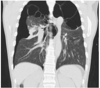
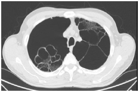
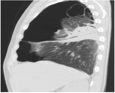
A 52-year-old male with a history of HIV infection and active smoking presented to the Emergency Room with right abdominal pain; he had no complaints of dyspnea or thoracalgia. On physical examination, he was tachycardic but eupneic and had no respiratory insufficiency; no breath sounds on both apexes and on the right hemithorax. Blood analysis was normal. Chest CT revealed an extensive right pneumothorax, partially septated by fibrotic bands between pleural leaflets; it also showed paraseptal emphysema with bigbullae (the biggest with 9 cm diameter).
Pneumothorax was drained and patient admitted for monitoring. He was discharged a few days later, but readmitted months after for another spontaneous pneumothorax.
Secondary pneumothorax is a complication of emphysema and its likelihood correlates with disease severity. It’s more frequent in males and in smokers. Abdominal pain is an atypical but possible presentation that can delay diagnosis and impair successful treatment.