
Journal of Clinical Images and Medical Case Reports
ISSN 2766-7820
Clinical Image - Open Access, Volume 4
Trypanosomes or platelet artefacts? A morphological dilemma!
Shruti Agrarwal1; Neha Singh2; B Priyavadhana3; Kriti Kaira4*
1Consultant Pathologist, Bansal Hospital, Bhopal 462016, India.
2Additional Professor, Department of Pathology and Laboratory Medicine, All India Institute of Medical Sciences, Rishikesh 249203, India.
3Assistant Professor, Department of Pathology and Laboratory Medicine, All India Institute of Medical Sciences, Rishikesh 249203, India.
4Post Doctoral Fellow in Oncopathology, Department of Pathology, Nizam’s Institute of Medical Sciences, Hyderabad 500082, India.
*Corresponding Author : Kriti Kaira, MBBS, MD
Post Doctoral Fellow in Oncopathology, Department of Pathology, Nizam’s Institute of Medical Sciences, Hyderabad 500082, India.
Ph: (+91) 9758031997;
Email: kaira.kriti@gmail.com
Received : Feb 02, 2023
Accepted : Feb 20, 2023
Published : Feb 27, 2023
Archived : www.jcimcr.org
Copyright : © Kaira K (2023).
Keywords: Peripheral smear; Platelets; Artefacts.
Abbreviations: PS: Peripheral Smear; K2-Edta: Dipotassium Ethylenediaminetetra acetic Acid.
Citation: Agrarwal AS, Singh N, Priyavadhana B, Kaira K, et al. Trypanosomes or platelet artefacts? A morphological dilemma! J Clin Images Med Case Rep. 2023; 4(2): 2303.
Description
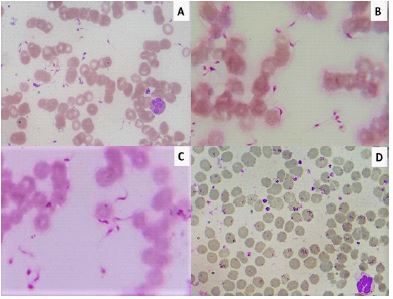
Peripheral Smear (PS) examination still remains a cornerstone and first step towards diagnostic work-up of patients. However, presence of artefacts in PS can sometimes prove perplexing for the pathologists [1]. In one such experience, early morning blood sample in K2-EDTA vial of a 23-year-old female was sent to hematology lab. Prepared PS revealed numerous extracellular, elongated, slender eosinophilic ‘flagellated’ structures measuring 4-5 μ, closely mimicking trypanosomes, but looking like miniature forms! With nondiscernible kinetoplasts (Figure 1A,B,C). Herhemoglobin was 11.5 g/dL, total leukocyte count 14.2 x 10⁹ /L and platelet count was 381 x 109 /L. She presented with abdominal distention, mild icterus and had mildly elevated total and direct bilirubin. There was no history of thrombocytopenia or fever and blood loss.
Three pathologists studied the smear, and their consensus that these structures were much smaller than the usual 10 μ size of trypanosomes [2], led to request for a repeat fresh blood sample. On repeat smear, these structures turned out to be normal round to oval platelets! (Figure 1D).
The patient was diagnosed eventually with choledochal cyst of liver with no evidence of any parasitic infection. Extensive smearing can cause platelet elongation, giving them a trypanosome-like appearance, which could be one of the contributing factors. Also, few studies suggested platelet morphogenesis takes place in plasma exhibiting platelets at various stages of maturation ranging from elongated proplatelets to curved fusiform disk shaped platelets which again can be mistaken for flagellated forms [3,4].
Various work on animal models stated platelets gets slightly activated by contact with the glass slide during smearing (as is common in feline samples) can acquire stellate form with dendritic processes [5].
According to Behnke’s hypothesis proplatelets appeared in plasmain a circadian rhythm, particularly during a few hours after sunrise in human blood. In this patient also, early morning sample showed elongated and spindled forms of platelets but repeat late morning sample was devoid of these flagellated forms [4].
Our observation emphasises the need for the pathologist to be aware of possible artefacts on PS. Each smear must be meticulously reviewed keeping in mind the clinical scenario and the limitation of the procedure, to avoid a misdiagnosis.
Declarations
Declaration of patient consent: The authors certify that they have obtained all appropriate patient consent forms.
Financial support and sponsorship: Nil.
Conflicts of interest: There are no conflicts of interest.
References
- Mc Namara C. Collection and handling of blood. In: Bain BJ, Bates I, Laffan MA, editors. Dacie and Lewis practical hematology. 12th ed. Edinburgh; Elsevier: 2017; 1-7.
- Bogitsh BJ, Carter CE, Oeltmann TN. Human parasitology. 5th ed. London; Elsevier: Chapter 6, Blood and tissue protozoa I: Hemoflagellates; 2018; 85-110.
- Wright JH. The histogenesis of the blood platelets J Morphol. 1910; 21: 263-278.
- Behnke O, Forer A. From megakaryocytes to platelets: Platelet morphogenesis takes place in the bloodstream. Eur J Haematol Suppl. 1998; 61: 3-23.
- Italiano JE Jr, Lecine P, Shivdasani RA, Hartwig JH. Blood platelets are assembled principally at the ends of proplatelet processes produced by differentiated megakaryocytes. J Cell Biol. 1999; 147: 1299‑312.