
Journal of Clinical Images and Medical Case Reports
ISSN 2766-7820
Clinical Image - Open Access, Volume 4
Malfunctioning tunneled dialysis catheter associated with arrhythmias
Mohankumar Doraiswamy1*; Khaled Boubes1; Nabil Haddad1; Anil Agarwal2
1Division of Nephrology, The Ohio State University, Columbus, OH, USA.
2VA Central California Health Care System, Fresno, CA, USA.
*Corresponding Author : Mohankumar Doraiswamy
Division of Nephrology, The Ohio State University, Columbus, OH, USA.
Ph: 615-315-1290, Fax: 614-293-3073;
Email: drmohankumar90@gmail.com
Received : Feb 02, 2023
Accepted : Feb 21, 2023
Published : Feb 28, 2023
Archived : www.jcimcr.org
Copyright : © Doraiswamy M (2023).
Keywords: Hemodialysis; Dialysis vascular access; Complications of tunneled catheter; Tunneled dialysis catheter; Arrhythmias on dialysis; Catheter malfunction.
Citation: Doraiswamy M, Boubes K, Haddad N, Agarwal A. Malfunctioning tunneled dialysis catheter associated with arrhythmias. J Clin Images Med Case Rep. 2023; 4(2): 2304.
Clinical image description
Tunneled Dialysis Catheters (TDC) remain a common modality of vascular access and often malfunction because of a myriad of reasons including thrombosis and fibrin sheath formation. A common practice is to use thrombolysis for TDC malfunctional with decreased blood flow. While this approach could work, it is prudent to obtain imaging of the TDC (cathetergram) to confirm its position and integrity. Here we show radiographic images of a right internal jugular TDC appropriately placed with the tip in the right atrium (Figure 1). Within two weeks, the patient developed poor blood flows on dialysis associated with intermittent arrhythmias. A Computed Tomography (CT) scan, done earlier that day to rule out pulmonary embolus, showed that the TDC had migrated medially (in wards) with the tip now positioned within the right ventricle (Figure 2). The TDC was exchanged, which resulted in the resolution of arrhythmias and excellent blood flow on dialysis. While medial migration of TDCs is rare, this case illustrates its possibility and the importance of imaging in evaluating TDC related complications. Failure to identify the cause of malfunction in this case not only would not have improved the blood flow, but moreover, could have resulted in serious complications such as arrhythmias, right ventricular rupture, or sudden cardiac death. Choosing proper length of TDC and location of exit site at the time of insertion is also important to prevent accidental migration into the right ventricle.
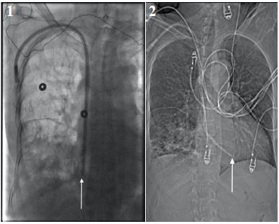
Figure 2: CT-scan imaging showing the TDC after it migrated medially with the tip in the right ventricle.
Declarations
Conflicts of interest: None.
Competing interests statement: The authors declare no conflicts of interest nor involved in promoting the therapies involved in the abstract.
Data sharing statement: Extra data is available by emailing corresponding author.
Acknowledgement: Not applicable.
Patient and public involvement statement: Patient accepted for publication. Public statement not applicable.
Cohort profile: Not applicable.
Funding disclosure: This research received no specific grant from any funding agency in the public, commercial or not-for-profit sectors. The authors declare no funding was received for this study.
Authors contributorship statement:
KB identified the case and helped in writing case presentation
MD, KB, AA and NH authored the manuscript
MD submitted the abstract to the Journal.
MD and KB helped in revision of the submission.
References
- Fiaccadori E, Gonzi G, Zambrelli P, Tortorella G. Cardiac arrhythmias during central venous catheter procedures in acute renal failure: A prospective study. J Am Soc Nephrol. 1996; 7: 1079-84.
- Agarwal AK, Haddad N, Boubes K. Avoiding problems in tunneled dialysis catheter placement. Semin Dial. 2019; 32: 535-540.
- Moist L, Vachharajani T. Vascular access for hemodialysis. Nissenson AR, Fine RN, eds. Handbook of Dialysis Therapy. 5th ed. Philadelphia, PA: Elsevier; 2016: 40-44.