
Journal of Clinical Images and Medical Case Reports
ISSN 2766-7820
Clinical Image - Open Access, Volume 4
Conjunctival inclusion cyst emerging from a pterygium
Yahya Saoiabi*; Hala El Belidi
Service d’ophtalmologie A de l’hôpital des spécialités, Université Mohammed-V-Souissi, Centre Hospitalier Universitaire, Rabat, Morocco.
*Corresponding Author : Yahya Saoiabi
Service d’ophtalmologie A de l’hôpital des spécialités, Université Mohammed-V-Souissi, Centre Hospitalier Universitaire, Rabat, Morocco.
Email: Saoiabi.yahya@gmail.com
Received : Feb 07, 2023
Accepted : Feb 22, 2023
Published : Mar 01, 2023
Archived : www.jcimcr.org
Copyright : © Saoiabi Y (2023).
Citation: Saoiabi Y, Belidi HE. Conjunctival inclusion cyst emerging from a pterygium. J Clin Images Med Case Rep. 2023; 4(3): 2306.
Case description
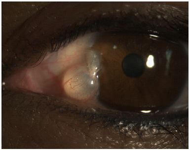
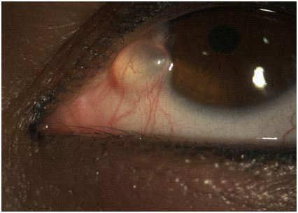
A 35 year-old female presented with a painless swelling of the left eye, with foreign body sensation, and an aesthetic concern.
There was no history of trauma or inflammatory episode. Ocular movements were normal.
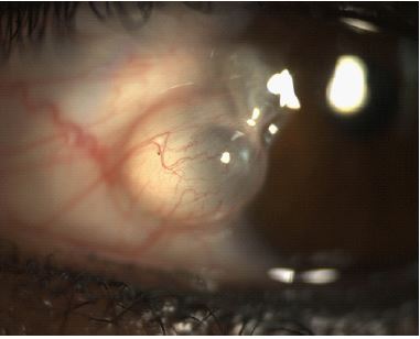
The blood vessels in the pterygium could be observed over the cyst, which is at the level of the body of the pterygium.
Conjuctival inclusion cysts are thin-walled benign cystic lesions, lined with a non-keratinizing epithelium containing serous fluid and slowly progressing cysts [1]. They are usually symptomless but can cause cosmetic disfigurement, reduced motility, foreign body sensation and dry eye due to unstable tear film when they increase in size [1].
Different etiologies, including epithelial implantation, parasitic, glandular retention, and lymphatic, can cause secondary conjunctival cysts [2]. Primary conjunctival inclusion cysts are congenital. Cysts in a pterygium can disrupt the tear film, cause dellen development, and indicate that the pterygium has to be removed [3].
References
- Mariana Nadais Aidar and al, Conjunctival Inclusion Cyst, eyewiki. 2022, https://eyewiki.aao.org/Conjunctival_Inclusion_Cyst
- Duke-Elder S, Leigh AG, Sys. of Ophtha. 1965; 3: 573.
- Kamel S, Bull. Ophthal .Soc. (Egypt). 1955; 48: 99.