
Journal of Clinical Images and Medical Case Reports
ISSN 2766-7820
Clinical Imaget - Open Access, Volume 4
Massive jugular foramen schwanoma revealed
by the hypogloss paralysis
Loudghiri Myriam1; Larhrabli Ibtissam1; Oukessou Youssef2; Rouadi Sami2; Redalah Larbi Abada2; Roubal Mohamed2; Mohammed Mahtar2
1ENT Head and Neck Surgery Department, Ibn Rochd University Hospital, Morocco.
2Faculty of Medicine and Pharmacy, Hassan II University, Casablanca, Morocco.
*Corresponding Author : Larhrabli Ibtissam
Residence Jnane Pasteur, Boulevard Abdelmoumen, Casablanca, Morocco.
Email: bettylar92@gmail.com
Received : Feb 03, 2023
Accepted : Feb 27, 2023
Published : Mar 06, 2023
Archived : www.jcimcr.org
Copyright : © Ibtissam L (2023).
Citation: Myriam L, Ibtissam L, Youssef O, Sami R, Larbi Abada R, et al. Massive jugular foramen schwanoma revealed by the hypogloss paralysis. J Clin Images Med Case Rep. 2023; 4(3): 2311.
Clinical image description
Jugular foramen schwannomas are a rare event with an estimated 200 cases reported in the literature. These tumors represent only 2.9% to 4% of all intracranial schwannomas and less than 1% of all temporal bone lesions [1,2].
In addition to the rarity of these tumors, their variable presentation, varying degrees of cranial nerve involvement, and multiple treatment options make each case unique. Signs and symptoms of patients often overlap with meningiomas, glomus jugular tumors, and occasionally vestibular schwannomas, making it difficult to differentiate preoperatively from jugular foramen schwannomas [3,4].
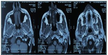
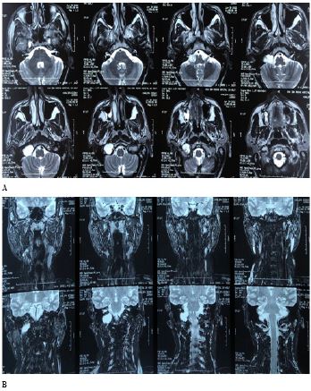
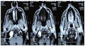
Modern imaging techniques are very helpful in diagnosis by distinguishing jugular foramen schwannomas from the more common jugular bulb. Computed Tomography (CT) and Magnetic Resonance Imaging (MRI) are often complementary and can show the extent of the tumor and also distinguish schwannomas from other tumors [5].
We report a case of a 55 years old man with no medical history presented to the consultation with a tongue’s right latero degation has been developing for a year. The physical examination revealed a paralysis of the right hypoglossal nerve without other associated signs, in particular nodysphonia or facial paralysis and no hearing loss. The MRI showed a 25 mm mass in the jugular foramen is what caused this one to expand. Regarding the extensive nature of the masse, the patient was referred to oncology for gamma knife radiotherapy.
References
- Tan LC, Bordi L, Symon L, Cheesman AD. Jugularforamen neuromas: A review of 14 cases. Surg Neurol. 1990; 34: 205–211.
- Samii M, Babu RP, Tatagiba M, Sepehrnia A. Surgical treatment of jugular foramen schwannomas. J Neurosurg. 1995; 82: 924–932.
- Lustig LR, Jackler RK. The variable relationship between the lower cranial nerves and jugular foramen tumors: Implications for neural preservation. Am J Otol. 1996; 17: 658–668.
- Jackson DG, Marzo S, Ishiyama A, Lambert PR. Glomus and Other Benign Tumors of the Temporal Bone. Philadelphia: Lippincott Williams & Williams. 2000.
- Fayad, Jose N, Keles, Bahar, Brackmann, Derald E. Jugular foramen tumors: Clinical characteristics and treatment outcomes. Otology & Neurotology. 2010; 31: 299-305.