
Journal of Clinical Images and Medical Case Reports
ISSN 2766-7820
Case Report - Open Access, Volume 4
Congenital duodenal windsock deformity simulating
midgut volvulus and treated with innovative minimal
invasive technique and duodenoplasty
Correia RC1*; Balliari MD1; Patel RV2
11Department of Pediatric Surgery and Neonatology, Unimed Hospital of Tatui, R. Cel. Lúcio Seabra, 960 - Centro, Tatuí - SP, 18270-240, Brazil.
22Departments of Pediatric Surgery, Postgraduate Institute of Child Health & Research and KT Children Govt University Teaching Hospital, Rajkot 360001, Gujarat, India.
*Corresponding Author : Rafael Cavalcante Correia
Pediatric Surgeon, Santa Casa de Tatui General Hospital, 3R. Cel. Lúcio Seabra, 960 - Centro, Tatuí - SP, 18270-240, Brazil.
Tel: +5515998106016;
Email: [email protected]
Received : Feb 14, 2023
Accepted : Mar 02, 2023
Published : Mar 09, 2023
Archived : www.jcimcr.org
Copyright : © Correia RC (2023).
Abstract
We present a 3200 gm female term neonate born by spontaneous vaginal delivery with uneventful antenatal scans and perinatal period who had passed normal meconium, breast fed for 6 days and presented with bilious vomiting simulating neonatal midgut volvulus. The baby was found to have double bubble in the upper abdomen and scattered gas bubble in the right lower abdomen on plain abdominal radiograph which was confirmed with abdominal ultrasound scan and an upper gastrointestinal contrast which showed duodenal obstruction at second part suggestive of windsock deformity. The patient underwent emergency exploratory laparotomy via minimal invasive periumbilical incision at which a partial duodenal obstruction with incomplete diaphragm with windsock deformity was seen and a vertical duodenotomy, resection of the duodenal diaphragm with small eccentric opening and transverse closure of the duodenotomy was carried out. The post-operative period was uneventful and at follow up the patient is thriving well.
Keywords: Acute abdomen; Bilious vomiting; Congenital; Duodenal diaphragm; Duodenal stenosis; Duodenoplasty; Midgut volvulus; Periumbilical laparotomy; Windsock deformity.
Citation: Correia RC, Balliari MD, Patel RV. Congenital duodenal windsock deformity simulating midgut volvulus and treated with innovative minimal invasive technique and duodenoplasty. J Clin Images Med Case Rep. 2023; 4(3): 2317.
Introduction
Congenital duodenal obstruction is more common with complete atresia in trisomy 21 and babies with VACTREL association and can be diagnosed prenatally as they have polyhydramnios and prenatal scan can easily diagnose them. Incomplete partial duodenal obstruction with fenestrated diaphragm with windsock deformity is a rare anomaly which can cause diagnostic challenges [1-6]. It commonly presents in the prenatal, neonatal period and early infancy with symptoms of partial obstruction but late presentations in childhood is well documented. A case of duodenal diaphragm with windsock deformity with normal prenatal scans, who has passed normal meconium with changing stools presenting acutely with bilious vomiting mimicking midgut volvulus and its accurate diagnosis with minimal invasive successful surgery is presented.
Case report
Term baby girl, weighing 3200 gm was born by spontaneous vaginal delivery with uneventful antenatal scans and perinatal period. The baby girl appeared normal at birth, discharged home the same day, had passed normal meconium and urine soon after birth and was breast feeding well with changing stools with no concerns. Suddenly the baby started bilious vomiting and upper abdominal fullness on the 6th day of life and stopped passing gas. The baby was immediately referred to us for further management. The baby was admitted in neonatal intestine care unit and resuscitated with intravenous fluid and nasogastric tube suction.
On examination baby was clinically well, active and slightly dehydrated. The abdomen was soft, compressible, not painful and had upper abdominal fullness distended. Rectal examination showed no grip on the examining finger, normal content and smear of stools and no gush of stools or gas after withdrawing the finger.
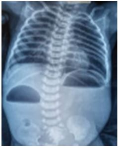
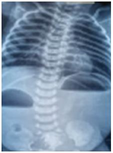
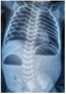
The laboratory investigations including blood tests and urine examination except for the blood gas analysis which showed mild acidosis. Plain abdominal radiograph demonstrated double bubble appearance in the upper abdomen with a bubble of gas in the right lower abdomen (Figure 1). Provisional differential diagnosis of neonatal midgut volvulus or congenital duodenal stenosis with diaphragm was made.
The abdominal ultrasound and color doppler studies confirmed dilated stomach and proximal duodenum with vigorous peristalsis and the orientation of superior mesenteric vessels appeared normal. An upper gastrointestinal contrast study was done to rule out midgut malrotation with volvulus which showed a dilatation of the stomach as well as proximal portion of the duodenum but without any passage of contrast to the distal duodenum or jejunum (Figure 2).
The baby underwent an emergency exploratory laparotomy via minimal invasive periumbilical cosmetic incision. At exploration, there were no signs of midgut malrotation and volvulus but a very dilated stomach and first portion of duodenum were observed. There was continuity of the duodenal wall externally and no signs of any complete atresia visible nor any external signs of a band externally to suggest the level of obstruction clearly at the junction of the dilated and collapsed duodenum but a slow transition was seen between the dilated and collapsed duodenum. The nasogastric tube was pushed manually which was tenting internally and could not be passed through the hole in the duodenal stenosis. It was possible to visualize the exact location of the membrane in the duodenum by careful inspection during surgery at this stage.
Duodenoplasty (Heine Mikulicz type) with resection of the duodenal diaphragm instead of Kimura’s diamond shaped duodenoduodenostomy was planned. A vertical lateral antimesenteric incision was done on this junctional part being tenting by the nasogastric tube and the lumen checked in the second portion of duodenum. It was possible to visualize the exact location of the membrane in the duodenum by careful inspection during surgery. A small orifice could be visualized in the duodenal diaphragm and a 4Fr nasogastric feeding tube was passed through it. This membrane around the small hole was grabbed with a tooth forceps and brought out through the duodenotomy (Figure 3). It wasn’t as thin as expected but an incision on the anterior to lateral part of the membrane was made to avoid inadvertent injury to the ampulla of Vater or the bile ducts medially, amplifying the lumen of the duodenum until a satisfactory aspect was achieved. A lateral duodenotomy with excision of the obstructive membrane was done for duodenal web and the duodenotomy was closed transversely using 5/0 interrupted delayed absorbable sutures in a single layer. The location of the web was between the first and second parts of duodenum. The duodenotomy was closed transversely to avoid any stenosis in the future.
Baby recovered very well, receiving feeds since the 2nd day post operatively, without vomiting or other concerns. She was discharged home after 5 days. At the follow up in clinics, she is asymptomatic, thriving well, eating and drinking normally and passing normal stools.
Discussion
The congenital incomplete duodenal stenosis with a diaphragm having an opening in it is very rare and may present at variable age with acute intestinal obstruction or can mimic volvulus or Hirschsprung’s disease [3]. Our case had no polyhy dramnios or associated anomalies like trisomy 21 or VACTREL association and was missed at prenatal scans. At birth baby had passed meconium immediately and was feeding and stooling well so Hirschsprung’s disease was unlikely.
In a large single centre series, duodenal atresia was the commonest cause of neonatal duodenal obstruction. Associated anomalies were noted in 61% patients, Down’s syndrome being the most frequent. These anomalies did not have any significant impact on the survival, nor did low birth weight. Preterm status had significant impact on prognosis [7].
In the current era, majority of cases are diagnosed in antenatal period but was missed in our case. It is very easy to diagnose the duodenal atresia antenatally due to complete obstruction as it leads to polyhydramnios in addition to dilated stomach and duodenum with double bubble or with associated anomalies like Trisomy 21 or VACTREL association duodenal obstruction is actively looked at due to high index of suspicion. Our case is a usual reminder of the fact that apparently normal looking babies at birth and feeding and opening bowel well may have an underlying partial intestinal obstruction like duodenal stenosis with a diaphragm and can easily be missed at birth and discharged home.
The clue to the existence of duodenal web with windsock deformity can be found at the plain radiograph with double bubble appearance and presence of bowel gas distally but midgut malrotation and volvulus needs to be excluded by an ultrasound with color doppler study and an upper gastrointestinal study as in our case. If there is no external clear indication of the transition and attachment of the membrane initially at exploration. Passage of a nasogastric tube manually may help locate the attachment and plan the duodenal incision.
Currently surgical exploration remains the gold standard for duodenal windsock deformity but in future with advancements in neonatal gastrointestinal endoscopy it may be possible to resect the web internally. We prefer minimal invasive exploration of the abdomen and the duodenum using periumbilical small cosmetic and minimal morbid incision and duodenoplasty rather than duodenoduodenostomy as it allows quick recovery and leads to minimal mortality or morbidity.
Duodenal diaphragm with a hole without any associated anomalies has better prognosis. Rattan et al had reported mortality of 11 (13.5%) out of 81 neonates [8], which is similar to the mortality rate in our group. Zamir et al had reported mortality of 58% in a series from Pakistan [9]. Escobar et al found 6% of late mortality attributed to associated anomalies, central nervous system bleeding, pneumonia, and anastomotic disruption etc. [10].
Conclusion
In conclusion, our case is an unusual and a rare presentation of an isolated congenital incomplete duodenal stenosis with a perforated diaphragm which illuded prenatal diagnosis and simulated neonatal midgut volvulus presenting at 6th postnatal day which was diagnosed accurately and treated successfully by minimal invasive surgery with periumbilical cosmetic mini-exploration, resection of the diaphragm and Heine Mikulicz type duodenoplasty rather than duodenoduodenostomy bypass with excellent prognosis.
References
- Patel RV, Kumar H, More B. Preampullary duodenal web simulating gastric outlet obstruction. J Neonat Surg. 2013; 2: 13.
- Patel RV. Congenital proximal jejunal diaphragm. JIMA. 1996; 94: 115-121.
- Patel R, Philip I: Distal duodenal stenosis in Down’s syndrome - a rare challenge. J Pediatr Surg Specialties. 2017; 11: 33-36.
- Patel RV, Govani D, Patel R, Dekiwadia DB. Bifid bile duct duodenal web bypass. Response to eLetter submitted to BMJ case reports by D Sfoungaris, Aristotelian University of Thessaloniki, Patel RV, Govani D, Patel R, Dekiwadia DB. Neonatal duodenoduodenostomy and missed duodenal stenosis with windsock deformity: a rare intraoperative error of technique and judgement by an unwary surgeon. BMJ Case Reports. 2014.
- Anthony FM, Chhaniara RA, Govani D, Corracia R, Gomes AL, et al. Congenital Neonatal Colonic Atresias arising in watershed areas of the colonic blood supply. JIMA.
- Patel RV, Govani D, Patel R, Dekiwadia DB. Neonatal duodenoduodenostomy and missed duodenal stenosis with windsock deformity: a rare intraoperative error of technique and judgement by an unwary surgeon. BMJ Case Rep. 2014.
- Kumar P, Kumar C, Pandey PR, Sarin YK. Congenital duodenal obstruction in neonates: Over 13 years’ experience from a single centre. J Neonat Surg. 2016; 5: 50.
- Rattan KN, Singh J, Dalal P. Neonatal duodenal obstruction: a 15-year experience. J Neonat Surg. 2016; 5: 13.
- Mirza B, Sheikh A. Multiple associated anomalies in patients of duodenal atresia: a case series. J Neonat Surg. 2012; 1: 23.
- Escobar MA, Ladd AP, Grosfeld JL, West KW, Rescorla FJ, et al. Duodenal atresia and stenosis: long term follow up over 30 years. J Pediatr Surg. 2004; 39: 867-871.