
Journal of Clinical Images and Medical Case Reports
ISSN 2766-7820
Short Report - Open Access, Volume 4
Congenital giant progressive parotid hemangioma in an infant treated successfully with propranolol and steroids
lamiGovani DJ1; Govani ND1; Govani DR1; Panchasara NG1; Patel RR1; Midha PK2; Swamy KB3; Patel RV1
1Departments of Pediatrics and Pediatric Surgery, Postgraduate Institute of Child Health & Research and KT Children Govt University Teaching Hospital, Rajkot 360001, Gujarat, India.
2J. Watumull Global Hospital & Research Centre, Delwara Road, Mount Abu, Rajasthan 307501, India affiliated to Medical Faculty of God Fatherly Spiritual University, Mount Abu, Rajasthan.
3Lincoln University College, Lincoln University, Kuala Lumpur, Malaysia.
*Corresponding Author : Ramnik Patel
Department of Pediatric Surgery, Postgraduate Institute of Child Health and Research and K T Children Government University Teaching Hospital Rajkot 360005 Gujarat India.
Tel : 00447956896641;
Email: ramnik@doctors.org.uk
Received : Feb 13, 2023
Accepted : Mar 02, 2023
Published : Mar 09, 2023
Archived : www.jcimcr.org
Copyright : © Patel R (2023).
Citation: Govani DJ, Govani ND, Govani DR, Panchasara NG, Patel RR, et al. Congenital giant progressive parotid hemangioma in an infant treated successfully with propranolol and steroids. J Clin Images Med Case Rep. 2023; 4(3): 2318.
Short report
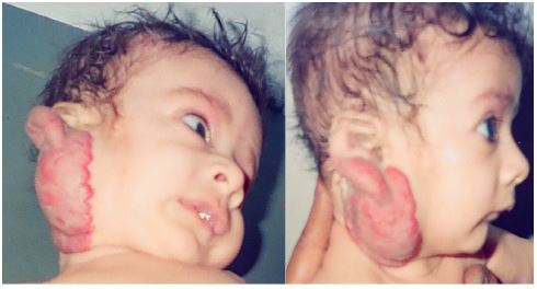
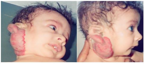
An otherwise healthy baby girl was born normally and a small birth mark like spot on her right parotid area appeared around two weeks of age. This spot went on growing gradually with growth of the body in infancy. However, the patient presented with cosmetically ugly giant mass pushing the right ear lobule up and compressing the right external ear canal with secondary changes started coming up with superficial ulceration and bleeding (Figure 1). There was no other lesion in the rest of the body. The patient was referred to ear, nose and throat/head and neck surgeon initially and surgery was advised. However, the parents were very poor and did not want an operation so visited us for second opinion. Color doppler ultrasonography confirmed the low flow state in the lesion consistent with the diagnosis of hemangioma. The oral propranolol was started at 1 mg/kg/day and titrated to 2 mg/kg/day with pulse rate and blood pressure monitoring for an intended duration of 12 months according to the Great Ormond Street Hospital protocol. Prednisolone was started at 0.3mg/kg/day and titrated to maximum dose of 3 mg/kg/day for 5 weeks in accordance to findings from a systematic review for the treatment of cutaneous haemangiomas [3,4]. The lesion started showing resolution while on therapy and At 3 month follow up, near complete resolution with some residual small residual pigmented scar noted (Figure 2).
Facial lesions are in infants and cosmetically disturbing [1] Vascular anomalies of the parotid gland are very rare and represent a spectrum of disorders from a simple “birthmark” to life- threatening entities simulating malignancy [2]. Hemangiomas have plump endothelia, increased mast cells, and multi-laminated basement membranes and are superficial, deep or combined and may be proliferating or involuting [3]. Accurate diagnosis is crucial for appropriate evaluation and management, although both types of simple vascular anomalies may appear similar apparently; there are several differences between them [4]. Treatment depends upon their size, location, and severity. Our case was treated with medical treatment with propranolol and systemic steroids. The medical treatment is very safe, effective, can be treated on outpatient basis, simple, low cost and gives excellent cosmetic and functional effects with improvement or complete resolution of symptoms and relief from the potential complications. The dose of prednisolone and its good effects for the treatment of infantile hemangioma is well documented in pediatric cases. The propranolol has less effects in older children with vascular malformations as compared to hemangiomas associated with higher failure rates and the side effects of hypoglycemia, sleepiness, GERD worsening, Irritable airways and bradycardia needs monitoring makes it a second choice as opposed to treatment of choice in young infants with proliferating lesions [5-8].
Declarations
Acknowledgments: We are grateful to Dr Jitendra G Govani, Primary Care Physician and Drs. Anilkumar and Kavita Trambadia MD, DCH consultant pediatrician for referring the patient to us and monitoring the systemic steroid therapy.
Conflict of interest: The authors have no conflict of interest to declare. No funding source was involved in this study.
Ethical approval: All procedures performed on human participants were in accordance with the ethical standards of the institutional and national research committee and with the 1964 Declaration of Helsinki and its later amendments or comparable ethical standards.
Informed consent: Informed consent was obtained from the parents and all the relatives involved prior to all the procedures. Parents and all involved parties were informed about the procedure.
References
- Patel RV, Gondalia JS. Congenital infiltrating lipomatosis of the face. British Journal of Plastic Surgery. 1991; 44: 157-158.
- Narasimharao KL, Patel RV, Mitra S, Bannerjee CK, Mitra SK. Mixed angioma of parotid gland simulating malignancy. Indian J Cancer. 1987; 24: 91-94.
- Patel R, Curry JI, Sinha CK, Davenport M. Chapter 62: Vascular Anomalies. In: Handbook of Pediatric Surgery. 2nd Ed, CK Sinha & Mark Davenport (Eds), Springer Verlag, London, 2021.
- Patel R, Curry JI. Vascular Malformations. In: Handbook of Pediatric Surgery. Part 4 Chap 13, 1st Ed, CK Sinha & Mark Davenport (Eds), Springer Verlag, London. 2010; 221-228.
- Patel RV, Govani DJ, Govani ND, Govani DR, Panchasara NG, et al. Giant cavernous vascular venous malformation of glans and distal penile shaft in a toddler treated successfully with steroids followed by circumcision. MedP J Cardiology and Vascular Medicine. 2022.
- Govani ND, Govani DR, Panchasara NG, Patel RR, Midha PK, et al. Unilateral congenital venous malformation in the form of giant right saphena varix and proximal varicose veins in a toddler boy. JIMA.
- Patel RV, Horton D, Robinson G, Lakshminarayan R, Daniel R. Congenital Bilateral Internal Jugular Venous Malformation in an Infant with Spinal Osteomyelitis and Discitis - Nature’s Recipe for Disaster at Vascular Access. J Pediatr Surg Specialities. 2016; 10: 28-33.
- Campbell A, Radford A, MacDonald A, Horton D, Besarovic S, et al. Phlebectasia of the Jejunum in a Child. J Pediatr Surg Specialities. 2015; 9: 35-39.