
Journal of Clinical Images and Medical Case Reports
ISSN 2766-7820
Case Report - Open Access, Volume 4
Paget’s disease of the inframammary fold
Verdonck Rikki1; Smeets Ann1; Nevelsteen Ines1; Floris Guiseppe2; Van Aerde Cedric2
1Multidisciplinary Breast Centre, University Hospital Gasthuisberg, Catholic University of Leuven, Belgium.
2Department of Imaging and Pathology, KU Leuven, Pathology, University Hospitals Leuven, Leuven, Belgium.
*Corresponding Author : Verdonck Rikki
Multidisciplinary Breast Centre, University Hospital Gasthuisberg, Catholic University of Leuven, Belgium.
Email: Rikki.Verdonck@uzleuven.be
Received : Feb 13, 2023
Accepted : Mar 10, 2023
Published : Mar 17, 2023
Archived : www.jcimcr.org
Copyright : © Rikki V (2023).
Abstract
Extramammary Paget’s disease is a rare malignancy of the skin. We present the case of a 62-year-old woman with a well-defined erythematous lesion of the left inframammary fold, without flaking, erosion or crust. Skin biopsy showed presence of Paget’s Disease. Physical examination and imaging showed no relation to the nipple-areola complex or breast glandular tissue. A wide local excision of the lesion was performed with 5 mm margins. Histology confirmed extramammary Paget’s disease. Adjuvant radiotherapy of the tumor bed was scheduled. In eczematous lesions not responding to topical treatment, a biopsy is needed for further diagnosis and to exclude Paget’s disease. Screening for underlying malignancy is guided by anatomical location. Although surgical treatment with clear margins is the golden standard, local recurrence rates remain high. Invasion into the dermis of >1 mm is the most important unfavorable prognostic factor.
Keywords: Extramammary Paget’s Disease; Diagnosis; Wide local excision; Case report.
Abbreviations: EMPD: Extramammary Paget’s Disease; MPD: Mammary Paget’s disease; WLE: Wide Local Excision ; DCIS: Ductal Carcinoma In Situ ; CEA: Carcinoembryonic Antigen; CK7: Cytokeratin 7; EMA: Epithelial Membrane Antibody; HER2: Membranous Human Epidermal Growth Factor Receptor 2; CK 5 /6: Cytokeratin 5/6; ER: Estrogen receptor; PR: Progesterone Receptor; GCDFP-15: Gross Cystic Disease Fluid Protein-15; MMS: Mohs Micrographic Surgery; SLNB: Sentinel Lymph Node Biopsy; RT: Radiotherapy.
Citation: MRikki V, Ann S, Ines N, Guiseppe F, Cedric VA. Paget’s disease of the inframammary fold. J Clin Images Med Case Rep. 2023; 4(3): 2329.
Introduction
Mammary Paget’s Disease (MPD) is defined as an in situ carcinoma of the nipple and areola. An underlying Ductal Carcinoma In Situ (DCIS) or invasive ductal carcinoma of the breast is present in >90% of the cases. Treatment is guided by the presence/absence of an underlying breast malignancy. Surgical treatment, mastectomy or breast conserving with nipple-areolar complex resection, is common practice [1]. Adjuvant endocrine and radiotherapy depends on performed surgery and underlying carcinoma. Extramammary Paget disease (EMPD) on the other hand is a rare adenocarcinoma of the skin and should be treated accordingly. EMPD presents as a slowly growing, erythematous or white plaque that mostly occurs on gland bearing areas of the skin. In Caucasian populations a female predominance is described, while in Asian populations male patients are predominant. It develops mostly in adults between 50-80 years, with a mean age of 65 years [2-4]. The pathogenesis is not completely understood [2,5]. We present a case of Paget’s disease located in the inframammary fold without any relation to the nipple or mammary glandular tissue.
Case report
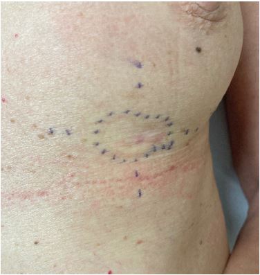
A healthy 62-year-old woman was referred to our consultation, requesting a second opinion. She complained of a red lesion in the left inframammary fold in the past year. Initially shaving and cryotherapy were performed by a dermatologist. Pathology showed M. Paget but the results were not discussed with the patient. Because the lesion persisted a skin biopsy was performed 6 months later. Paget’s disease was confirmed and she was referred to a gynecologist. There was no evidence of intramammary lesions on mammography, neither by breast ultrasound. Follow-up was proposed with imaging after 6 months. However, the patient was not comfortable waiting another 6 months and made an appointment in our center. Clinical examination showed a skin lesion in the medial part of the left inframammary fold of approximately 20 mm by 30 mm (Figure 1). It was slightly erythematous, flat and had well-defined margins. There was no crust or erosion and there were no scales. Thorough clinical examination of the breasts revealed no other abnormalities, especially not towards the nipple-areola complex. There was no evidence of a supernumerary nipple, nor ectopic breast tissue. The tissue samples were requested for revision and additional tomosynthesis and MRI of the breasts were planned to exclude underlying intramammary pathology. Pathology review of the skin biopsy at our center confirmed presence of Paget’s disease, additional imaging excluded intramammary lesions. The diagnosis of extramammary Paget’s disease was proposed. We performed a wide local excision of the lesion with a macroscopic 5 mm margin. The defect was primarily closed by our reconstructive surgeons (Figure 2). There was an uneventful recovery.
Histopathological examination showed either clustered polygonal cells or scattered isolated malignant cells with relatively abundant pale cytoplasm in the epidermis. The nuclei were enlarged, vesicular with irregular nuclear borders and hyperchromatic. There was no evidence of invasion in the dermis. Underlying glandular structures without any connection to the intraepithelial lesion were noticed as well. Immunohistochemical analysis showed a positive staining for Carcinoembryonic Antigen (CEA), Cytokeratin 7 (CK7), Epithelial Membrane Antibody (EMA) and strong membranous human epidermal growth factor receptor 2 (HER2) staining that was noticeable at low power magnification. Cytokeratin 5/6 (CK 5 /6), Melan A and S100 were negative. Estrogen receptor (ER) showed moderate nuclear expression in 15% of the intraepidermal malignant cells, progesterone receptor (PR) was negative. The diagnosis of intra-epithelial extramammary Paget’s disease was confirmed after exclusion of melanoma in situ, spinocellular carcinoma in situ and Toker’s cell hyperplasia.
At our multidisciplinary meeting adjuvant radiotherapy of the tumor bed was proposed consisting of 16 sessions with 2.66 Gy per fraction. Radiotherapy was started 6 weeks after surgery. Considering EMPD as a primary skin tumor, no adjuvant endocrine therapy was suggested.
Discussion
Clinical presentation
Most commonly patients present with a well-described, slowly growing, asymmetrical, erythematous or white lesion. Crust, scales and erosion can also be present, potentially mimicking other skin disorders. Associated symptoms such as pruritus (most common), tenderness, burning, irritation, bleeding and swelling are present in 90% of the patients. Benign/reactive local lymph node enlargement is possible [2,4,5]. Due to a wide range of symptoms and variable clinical aspects, misdiagnosis is common. Differential diagnosis includes contact dermatitis, melanoma, psoriasis, eczema, lichen sclerosus , fungal infection and mycosis fungoides [5]. Genital, perianal and pubic area involvement is most common. However, trunk, perioral skin, oral mucosa, scalp, axillary or umbilical regions can also be affected [3,4]. Dermoscopy is a non-invasive technique that can contribute to the clinical diagnosis. Most dermoscopic findings are similar to other skin disorders in the differential diagnosis, although milky-red areas are significantly more prevalent in EMPD than in eczema, fungal infection or Bowen disease. Vascular structures are also more present in EMPD than in eczema or fungal infection [6].
To our knowledge, this is the first case report of a lesion in the inframammary fold.
Histology
When standard topical treatment fails, skin biopsy is recommended in all patients with pruritic eczematous lesions in apocrine gland-bearing locations [2,5,7,8]. Typically, intraepidermal clusters of pale staining cells, larger than keratinocytes are seen. The cells contain pleomorphic nuclei with pronounced atypia, prominent nucleoli and mitotic figures [8]. EMPD can be divided into 2 types: primary and secondary. Primary EMPD is an intraepithelial neoplasm of the epidermis, where invasion can develop over time. Primary EMPD can be further subdivided in epidermal EMPD (in situ carcinoma) and invasive EMPD (invasion into the dermis or even deeper). Secondary EMPD on the other hand, is defined by epidermotropic spread of malignant cells or direct extension in the epidermis from an underlying adenocarcinoma (e.g., colorectal or urothelial carcinoma) [2,4]. Immunohistochemical staining can be used to guide the differential diagnosis. The panel should include CK7, CK20, CEA, EMA, HER2 and gross cystic disease fluid protein-15 (GCDFP-15). Primary EMPD is typically CK7-positive, CK20-negative and GCDFP-15-positive. Secondary EMPD is obviously more heterogeneous. HER2 and androgen receptor expression are frequently found. In 12% of the cases, ER is positive and only 9% express PR [9]. The most common underlying malignancies are colorectal carcinoma (CK7-negative, CK20-positive, GCDFP-15-negative) and urothelial carcinoma (CK7-positive, CK20-positive, GCDFP-15-negative). Tissue-specific antigens can be used for further differentiation, but careful clinicopathological correlation is crucial [2,4,7]. In the differential diagnosis spinocellular carcinoma in situ and melanoma in situ should be included as well. Smart use of IHC in combination with PAS-staining are generally sufficient to exclude these entities. In our case, given the location and the possible relation with the milk line we also wanted to exclude Tocker’s cell hyperplasia by combining CEA with above mentioned immunostainings.
Diagnostic work-up
Underlying associated malignancy is reported in 7-40% of the EMPD [2], therefore further diagnostic work-up is recommended upon histologic diagnosis. A comprehensive anamnesis and thorough clinical examination always need to be performed [2,5,7]. Further imaging and fine needle aspiration cytology ought to be used to investigate palpable lymph nodes [7]. The anatomical region of involvement should guide test selection, e.g.: colposcopy and urine cytologic screening for vulvar EMPD, or anoscopy and colonoscopy for perianal EPMD [7]. Schmitt et al. proposed a screening algorithm for all patients that included age-appropriate cancer screening, urine cytology, mammography for women and prostate specific antigen (PSA) blood test for men [10]. We performed extensive mammary imaging in our case to exclude intramammary pathology. No further (distant) screening was conducted. Currently, no validated TNM staging system for EMPD exists.
Management
Surgical resection is the golden standard as primary treatment. However, because EMPD is mostly seen in regions where wide local excision (WLE) implies mutilating surgery, the extent of the excision should always be individualized [7]. Reported recurrence rates after WLE are high, between 30% and 60% [3,7,11]. In literature, there is no consensus on margin size. A 1 cm margin for clinically well-defined lesions and a 2 cm margin in ill-defined lesions is considered safe [4,5,11,12]. Some reports advocate for even wider excision with clinical margins ranging up to 5 cm. However, there is no association between margin size and risk of recurrence [3,11]. Mapping biopsies are not required for well-defined lesions or when 2 cm margins can be achieved [4,11,12]. Mohs micrographic surgery (MMS) is expensive and time-consuming but may lower recurrence rates. In comparison to other nonmelanoma skin cancers, the recurrence rate of 12,2% in EMPD after MMS is still relatively high [13]. Possible explanations can be that it is difficult to identify EMPD microscopically in frozen sections and the multifocal nature of the disease. Several ways to aid identification have been described, e.g.: quality staining, thinner sections, slow Mohs and especially intraoperative use of immunohistochemistry (CK7 and CEA) [7,8,11].
Several studies evaluated the use of sentinel lymph node biopsy (SLNB). None of the patients with intraepthelial EMPD had a positive SLNB [14]. Positivity rates were 15 and 16,9% in patients with invasive EMPD and clinical negative nodes [14,15]. Because the positivity ratio in invasive EMPD is quite high, the use of SLNB can be considered. However, one should bear in mind that it is unclear whether disease free survival is improved by lymph node dissection in SLNB-positive cases. No consensus exists for lymph node dissection or targeted removal of affected nodes. There is also no clear evidence that overall survival is improved by lymph node dissection or targeted lymph node dissection [7]. When morbidity of surgery is too high, other non-invasive treatments can be considered.
Primary treatment with Imiquimod 5% cream has been used in EMPD. Complete response rates between 30 and 56% [4,7,11,16] and recurrence rates up to 35,4% [7,16] are found in literature. Long term efficacy is unclear due to short duration of follow-up [7,8]. Neoadjuvant treatment with Imiquimod may be useful, although a higher recurrence rate after surgery was demonstrated in one study [4,17].
Radiotherapy (RT) can be used as primary or adjuvant treatment. In primary treatment a margin of 3,5 cm from the clinical border is advisable, if possible [7]. In patients with primary EMPD and contraindications for surgery, use of RT as definitive treatment is feasible. Adjuvant RT also seems useful in patients with positive or close margins [18]. In patients with dermal invasion, treatment of regional lymph nodes is advised, although no clear evidence of benefit exists [7,18]. When there is dermal invasion or close margins a dose of 45 to 60 Gy should be enough for local control and to prevent lymph node invasion. A dose of 60 Gy is advised in patients with enlarged lymph nodes or positive margins [18].
Overall, data suggest that photodynamic therapy shouldn’t be used with a curative intent but symptom reduction in a palliative setting is possible [4].
HER2 targeted therapies in monotherapy or in combination with chemotherapy is a treatment option in HER2-positive advanced EMPD [4,5,9]. A recent meta-analysis showed HER2 expression in 32% of female patients and 26% of male patients. A possible positive correlation is present between HER2 overexpression and disease recurrence, lymph node metastasis and dermal invasion. In contrast to described discordance rates of HER2 expression between primary tumor and distant metastasis in breast and gastric cancer, there is a good overall concordance in EMPD [9].
Data on endocrine receptors are limited. While ER and PR expression is rather low, androgen receptor (AR) is present in almost half of the patients with EMPD. So anti-androgen therapy can be promising but further research is necessary [9].
For metastatic EMPD, chemotherapy can be considered. Multiple treatment regiments are reported, but no consistent data exist. Possible used regiments are low-dose-5-fluorouracil/cisplatinin, docetaxel monotherapy, FECOM (5-fluorouracil, epirubicin, carboplatin, vincristine and mitomycin C), PET (cisplatin, epirubicin and paclitaxel), S-1 monotherapy and S-1 combined with docetaxel. No complete response is obtained but quality of life can often be improved [4,7,8,11].
Follow-up
No validated algorithm is available for follow-up after treatment for EMPD. Appropriate follow-up is based on clinical judgement of the physician. Physical examination with lymph node examination is advised every 3 to 6 months during the first 3 years and every 6 to 12 months until at least 5 years after diagnosis. Screening for associated malignancies and metastasis guided by anatomical location may be considered in EMPD [7].
Prognosis
Primary EMPD originates as an in situ carcinoma and has no risk of metastasis [10]. However, over time primary EMPD can invade the dermis. Invasive EMPD does have the potential of lymphovascular invasion and metastases. There’s a minimal risk of nodal involvement when invasion is < 1 mm from the dermoepidermal junction. Invasion of >1 mm into the dermis can b
Conclusion
EMPD is a rare cutaneous adenocarcinoma. Delayed diagnosis is frequent due to the clinical resemblance to more common skin disorders. When standard topical treatment fails, a biopsy is crucial. Screening for underlying malignancy is advised and should be individualized. Treatment is focused on wide local excision with histologic clear margins if possible. Sentinel lymph node biopsy, lymph node dissection or adjuvant therapy are no clinical routine and should always be individualized. Non-surgical treatment can be considered when surgery is not feasible. Prognosis depends on the level of invasion into the dermis, with invasion >1 mm as the most important unfavorable prognostic factor.
References
- Markarian S, Holmes DR. Mammary Paget’s Disease: An Update. Cancers (Basel). 2022; 14.
- Morris CR, Hurst EA. Extramammary Paget Disease: A Review of the Literature—Part I: History, Epidemiology, Pathogenesis, Presentation, Histopathology, and Diagnostic Work-up. Dermatologic Surgery. 2020; 46: 151-158.
- Wollina U, Goldman A, Bieneck A, Abdel-Naser MB, Petersen S. Surgical Treatment for Extramammary Paget’s Disease. Curr Treat Options Oncol. 2018; 19: 27.
- Ishizuki S, Nakamura Y. Extramammary Paget’s Disease: Diagnosis, Pathogenesis, and Treatment with Focus on Recent Developments. Curr Oncol. 2021; 28: 2969-2986.
- Adashek JJ, Leonard A, Nealon SW, Krishnan A, Mosiello GC, et al. Extramammary Paget’s disease: what do we know and how do we treat? Can J Urol. 2019; 26: 10012-10021.
- Mun JH, Park SM, Kim GW, Song M, Kim HS, et al. Clinical and dermoscopic characteristics of extramammary Paget disease: a study of 35 cases. Br J Dermatol. 2016; 174: 1104-1107.
- Kibbi N, Owen JL, Worley B, Wang JX, Harikumar V, et al. Evidence-Based Clinical Practice Guidelines for Extramammary Paget Disease. JAMA Oncol. 2022; 8: 618-628.
- Merritt BG, Degesys CA, Brodland DG. Extramammary Paget Disease. Dermatol Clin. 2019; 37: 261-267.
- Angelico G, Santoro A, Inzani F, Straccia P, Arciuolo D, et al. Hormonal Environment and HER2 Status in Extra-Mammary Paget’s Disease (eMPD): A Systematic Literature Review and Meta-Analysis with Clinical Considerations. Diagnostics. 2020; 10: 1040.
- Schmitt AR, Long BJ, Weaver AL, McGree ME, Bakkum-Gamez JN, Brewer JD, et al. Evidence-Based Screening Recommendations for Occult Cancers in the Setting of Newly Diagnosed Extramammary Paget Disease. Mayo Clin Proc. 2018; 93: 877-883.
- Morris CR, Hurst EA. Extramammary Paget’s Disease: A Review of the Literature Part II: Treatment and Prognosis. Dermatologic Surgery. 2020; 46: 305-311.
- Kaku-Ito Y, Ito T, Tsuji G, Nakahara T, Hagihara A, Furue M, et al. Evaluation of mapping biopsies for extramammary Paget disease: A retrospective study. J Am Acad Dermatol. 2018; 78: 1171-1177.e4.
- Bae JM, Choi YY, Kim H, Oh BH, Roh MR, Nam K, et al. Mohs micrographic surgery for extramammary Paget disease: a pooled analysis of individual patient data. J Am Acad Dermatol. 2013; 68: 632-637.
- Ogata D, Kiyohara Y, Yoshikawa S, Tsuchida T. Usefulness of sentinel lymph node biopsy for prognostic prediction in extramammary Paget’s disease. Eur J Dermatol. 2016; 26: 254-259.
- Fujisawa Y, Yoshino K, Kiyohara Y, Kadono T, Murata Y, et al. The role of sentinel lymph node biopsy in the management of invasive extramammary Paget’s disease: Multi-center, retrospective study of 151 patients. J Dermatol Sci. 2015; 79: 38-42.
- van der Linden M, van Hees CL, van Beurden M, Bulten J, van Dorst EB, et al. The Paget Trial: topical 5% imiquimod cream for noninvasive vulvar Paget disease. Am J Obstet Gynecol. 2022; 227: 250.e1-250.e8.
- Choi S, Oh Y, Roh MR, Chung KY, Oh BH. Initial topical monotherapy may increase the risk of recurrence in patients with extramammary Paget’s disease. J Dermatol. 2021; 48: 585-591.
- Tagliaferri L, Casà C, Macchia G, Pesce A, Garganese G, et al. The Role of Radiotherapy in Extramammary Paget Disease: A Systematic Review. International Journal of Gynecologic Cancer. 2018; 28: 829-839.
- Karam A, Dorigo O. Treatment outcomes in a large cohort of patients with invasive Extramammary Paget’s disease. Gynecol Oncol. 2012; 125: 346-351.