
Journal of Clinical Images and Medical Case Reports
ISSN 2766-7820
Short Report - Open Access, Volume 4
Hypothyroidism presenting with left sided Brown’s syndrome
Akhil Shah1; Lesley Kaye1; Sze May Ng1,2*
1Paediatric Department, Southport and Ormskirk NHS Trust, Ormskirk, UK.
2Department of Women’s and Children’s Health, University of Liverpool, Liverpool, UK.
*Corresponding Author : Sze May Ng
Paediatric Department, Southport and Ormskirk NHS Trust, Ormskirk, UK.
Email: may.ng@nhs.net
Received : Feb 27, 2023
Accepted : Mar 15, 2023
Published : Mar 22, 2023
Archived : www.jcimcr.org
Copyright : © Ng MS (2023).
Abstract
An 11-year-old female presents with a 6-week history of double and blurred vision associated with headaches and neck swelling. Thyroid function tests demonstrated antibody negative hypothyroidism. In addition, connective tissue disorder screening was negative. The patient was commenced on thyroid hormone replacement (levothyroxine).
However, 5 days later, she re-presented with strabismus and progressive diplopia. Intracranial imaging was performed to rule out space occupying masses. A diagnosis of left sided Brown’s syndrome secondary to hypothyroidism was made. Initial treatment consisted of conservative management with a Fresnel prism whilst continuing levothyroxine to reach euthyroid status. Patient has remained clinically stable since presentation with no further recurrence of symptoms. There are no known previously documented cases in the current literature.
Citation: Shah A, Kaye L, May Ng S. Hypothyroidism Presenting with Left Sided Brown’s Syndrome. J Clin Images Med Case Rep. 2023; 4(3): 2335.
Background
Brown’s syndrome is characterised by the inability to elevate the affected eye in adduction due to a mechanical restriction of the superior oblique muscle tendon. Most commonly a congenital condition, acquired cases are usually caused by surgery, trauma or disorders associated with inflammation such as sinusitis, system lupus erythematosus or juvenile rheumatoid arthritis [1,2]. This is the first published case where Brown’s syndrome formed part of the clinical presentation of antibody negative hypothyroidism.
Case presentation
An 11 year old female was referred after presenting to her general practitioner with a 6 week history of intermittent double vision with blurring, headaches, fatigue and feeling cold. Laboratory investigations revealed raised levels of thyroid stimulating hormone and insufficient free thyroxine (T4) demonstrating primary hypothyroidism. She was also found to have neck swelling and dry skin. There were no abnormalities on eye examination at this stage. The child had a history of autoimmune thrombocytopaenia that required treatment with intravenous immunoglobulins but had resolved 7 years earlier. She was commenced on thyroid hormone replacement (levothyroxine) whilst awaiting imaging and follow-up.
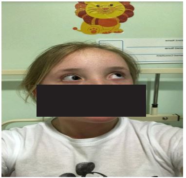
After 5 days, the patient re-presented due to a new onset squint of her left eye associated with a “popping” sensation and worsening diplopia. She was noted to have asymmetric eye movements with inability to elevate her left eye on right lateral gaze (adduction). There was no evidence of ptosis, proptosis or other abnormalities typically associated with thyroid disease. She underwent ophthalmology assessment and was diagnosed with left Brown’s syndrome secondary to thyroid disease (Figure 1).
Investigations
Further tests after her first presentation included thyroid antibody and connective tissue disorder screening, an adrenocorticotrophin stimulating test and glucose levels which were all normal. Importantly, anti-thyroid peroxidase antibodies were not detected. An ultrasound scan described findings consistent with a diffuse goitre. Computerised tomography performed at the time of her second presentation and a follow-up magnetic resonance image of the brain was reported as normal with no evidence of a mass lesion. Specifically, there were normal appearances of the extra-ocular muscles.
A repeat thyroid ultrasound 3 years 8 months from first presentation demonstrated a mildly enlarged thyroid gland with a heterogenous texture and increased vascularity consistent with thyroiditis.
Differential diagnosis
Intracranial mass
Connective tissue disorder
Cranial nerve palsy
Treatment
The patient received thyroid hormone replacement as levothyroxine, currently at 87.5 micrograms once daily. The resultant squint was treated conservatively with a Fresnel prism until resolution of her squint once she reached euthyroid status.
Outcome and follow-up
The patient received ophthalmology follow-up for 1 year. She has remained under follow-up in the paediatric endocrinology clinic for 4 years and 11 months. She has remained clinically stable with dosage adjustments according to serial blood test monitoring. There has been no recurrence of symptoms.
Discussion
Extra-ocular muscle involvement is well-recognised in hyperthyroid states, such as Grave’s disease, and is commonly known as thyroid eye disease. This is a restrictive problem largely involving the recti muscles, most commonly the inferior rectus [3,4]. Superior oblique muscle involvement has been proposed to be more common that previously recognised in thyroid eye disease [3,5]. There are also previous reports of Brown’s syndrome possibly due to thyroid eye disease [3]. However, there have been no previously documented cases secondary to hypothyroidism. This should be an important differential in the presentation of Brown’s syndrome.
Learning points
o Most cases of Brown’s syndrome are congenital
o Acquired cases are usually secondary to inflammatory conditions
o Extra-ocular muscle pathology is a recognised consequence of thyroid eye disease, with superior oblique enlargement becoming more evident.
o There are no previously published cases of Brown’s syndrome secondary to hypothyroidism.
o Thyroid disease could be an important differential diagnosis to consider with the presentation of Brown’s syndrome.
References
- Brown Syndrome - American Association for Pediatric Ophthalmology and Strabismus.
- Brown Syndrome - NORD (National Organization for Rare Disorders).
- Thacker NM, Velez FG, Demer JL, Rosenbaum AL. Superior oblique muscle involvement in thyroid ophthalmopathy. J AAPOS. 2005; 9: 174-178.
- Bahn RS. Graves’ ophthalmopathy. N Engl J Med. 2010; 362: 726-738.
- del Porto L, Hinds AM, Raoof N, Barras C, Davagnanam I, et al. Superior oblique enlargement in thyroid eye disease. J AAPOS. 2019; 23: 252.e1-252.e4.