
Journal of Clinical Images and Medical Case Reports
ISSN 2766-7820
Review Article - Open Access, Volume 4
Right ventricular dysfunction in cancer patients; Is it
real or dilemma? A narrative review
Azin Alizadehasl1; Elgar Enamzadeh1; Alia Bahramnejad1; Hanie Hajiali Fini2; Rezvaneh Shourmeij1; Amir Dousti3; Mina Mohseni1*; Kamran Roudini4; Aghdas Shadmehr2
1Cardio-Oncology Research Center, Rajaie Cardiovascular Medical and Research Center, Iran University of Medical Sciences, Tehran, Iran.
2Echocardiography Research Center, Rajaie Cardiovascular Medical and Research Center, Iran University of Medical Sciences, Tehran, Iran.
3Interventional Cardiology Research Center, Rajaie Cardiovascular Medical and Research Center, Iran University of Medical Sciences, Tehran, Iran.
4Cancer Institute, Imam Khomeini Hospital, Tehran University of Medical Sciences, Tehran, Iran.
*Corresponding Author : Mina Mohseni
Cardio-Oncology Research Center, Rajaie Cardiovascular Medical and Research Center, Iran University of Medical Sciences, Tehran Iran.
Email: cardiooncology.r@gmail.com
Received : Feb 27, 2023
Accepted : Mar 15, 2023
Published : Mar 22, 2023
Archived : www.jcimcr.org
Copyright : © Mohseni M (2023).
Abstract
Cardiotoxicity is one of the challenges of treating a cancer patient. With the advancement of treatment methods and the production of new drugs, the life expectancy of cancer patients improved significantly. As a result, cardiovascular diseases that may occur due to cancer treatment need special attention. In most studies, only the function of the left ventricle is evaluated to investigate cardiotoxicity, while the right ventricle can also be affected by anticancer treatments. Right ventricular dysfunction can help predict the occurrence of left ventricular dysfunction. In addition, it can be the cause of many cardiovascular symptoms of the patient and even affect cardiovascular mortality in cancer patients. The methods of diagnosing right ventricular dysfunction have made many advances, including advanced echocardiography and cardiac MRI methods. In this review, we examined studies that are about right ventricular function and methods of diagnosing right ventricular dysfunction in cancer patients.
Citation: Alizadehasl A, Enamzadeh E, Bahramnejad A, Fini HH, Mohseni M, et al. Right ventricular dysfunction in cancer patients; Is it real or dilemma?. A narrative review. J Clin Images Med Case Rep. 2023; 4(3): 2336.
Background
With the advancement of cancer treatment drugs, the life expectancy of cancer patients has increased. Both due to the development of drugs and the production of new drugs and due to the increased survival of patients, attention should always be paid to unwanted side effects caused by treatment, especially cardiovascular side effects. The most important traditional chemotherapy drugs that cause cardiotoxicity are drugs of the anthracycline group. Other drugs including Her2 inhibitors, proteasome inhibitors, platinum-based chemotherapeutic drugs, antimetabolites, vascular endothelial growth factor inhibitors, microtubule inhibitors, thyrosine kinase inhibitors, targeted agents and immunotherapies can also have cardiovascular side effects [1,2].
The most common cardiovascular side effects of these drugs can be Left Ventricular (LV) dysfunction, heart failure, hypertension, cardiac arrhythmia, ischemic heart disease and vascular events [2].
In the follow-up and investigation of cardiac complications of anti-cancer treatments, usually only attention is paid to the function of the LV [3]. While the right ventricle (RV) can also be impaired, and the cause of the patient’s symptoms (e.g. dyspnea) may be the dysfunction of the RV [4].
In this review, we decided to examine the studies on RV function in cancer patients, diagnostic methods, and the most important anticancer drugs that cause RV dysfunction.
Method
In this review, all documents in the field of right ventricular function of cancer patients undergoing chemotherapy were considered as the study population of this research. Search using keywords such as: cancer, chemotherapy, anthracycline, right ventricle, echocardiography and global longitudinal strain in relevant databases, including Medline, Scopus, Embase, Cochrane central, register of controlled trials and the Cochrane database of systematic reviews (search for word combinations). All relevant descriptive, interventional, clinical trials, case-control or cohort studies, and case reports were included in this review. Reference lists of relevant articles found were also reviewed to identify other relevant studies. The search procedures were as follows: 1) First, the words cancer and chemotherapy were searched. 2) The right ventricle was also added. 3) Then, based on MeSH words, advanced chemotherapy and echocardiography words were added. All found articles were studied and summarized and written in a categorized form.
RV function
The importance of right ventricle (RV) as a predictor of prognosis and symptom occurrence was neglected for several decades [5]. Evaluation of RV is a main part of echocardiography. However, it remains challenging due to the complex anatomy of the RV. Visual assessment is the simplest and the most common method for RV function evaluation. Although, echocardiography guidelines suggest at least one quantitative parameter in addition to it including tricuspid annular plane systolic excursion (TAPSE), tissue Doppler imaging at base of RV lateral wall (RV S’), longitudinal strain of free lateral wall of RV, RV fraction area change (RVFAC) and three dimensional estimation of RV function [6] (Figure 1).
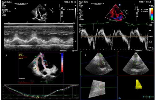
TAPSE reveals longitudinal RV contractility (movement of the lateral part of the TV annulus toward RV apex). It is assessed by M-mode from apical 4 chamber view in Transthoracic Echocardiography (TTE) and is one of the useful methods because of simplicity and lower dependency to optimal imaging quality [7]. Velocity of tricuspid annulus longitudinal motion is evaluated by tissue Doppler as RV S’ or systolic excursion velocity. RV S’ is more accurate than FAC and TAPSE when RVEF by CMR is normal or mildly abnormal. The longitudinal strain of the RV free wall is also a method of RV assessment and measured by speckle tracking imaging in apical 4 chamber view. Cut off of -20% correlates with normal RV function. Some studies reveal this parameter can predict early stages of RV dysfunction [8]. FAC is a common parameter for RV function assessment and reflects the volume change during RV systolic and diastolic phases based on 2D measurements. RV FAC less than 35% represents RV dysfunction [6,8]. Eventually, due to RV structural complexity, 3D echocardiography is currently used as an accurate and reliable method for RV function evaluation compared to above 2D based methods. Studies showed strong correlation between 3D echocardiography and CMR RV volume and function [9].
Traditional echocardiography and RV function in cancer patients
In traditional 2D echocardiography in patients with breast cancer treated with anthracyclines, RV end-diastolic diameters and the index, RVFAC and TAPSE were measured and a significant decrease in RV systolic and diastolic function occurred during treatment with anthracyclines, although in Esfahani et.al study they were in the normal range. This reduction can be a predictor of cardiotoxicity in the future [10]. The amount of change in RVFAC and TAPSE between before the onset of the chemotherapeutic regimen (T1) and after the completion of two cures of the regimen (T3) were found to be correlated with the amount of change in NT-proBNP measurements between T1 and T3. subclinical decrease in right ventricular systolic and diastolic echocardiographic indices occurs in a relatively short time interval after onset of chemotherapy [11].
Advanced echocardiography and RV function in cancer patients
In some studies, RV myocardial injury occurs simultaneously with LV cardiotoxicity by using longitudinal strain with similar cutoff percent change [12]. But some studies with advanced echocardiography suggest that the Right ventricular global longitudinal strain (RV-GLS) may be more vulnerable than the left ventricle to anthracycline therapy. Significant reduction is seen in RV GLS and RV free wall longitudinal strain (RV-FWLS) systolic peak in breast cancer patients who were treated with anthracycline [13]. Endocardial and mid-myocardial layer strains of RV are more affected than epicardial strain in the cancer patients. It is interesting that decreased in RV longitudinal strain associated with The presence of cancer [14]. Compared to traditional parameters, RV Speckle Tracking Echocardiography (STE) and 3D echocardiography appeared to detect anthracycline-related subclinical damage earlier [15]. RV 2D speckle-tracking echocardiography is the best modality for detecting early impairment of RV function compared to traditional modalities [16]. RV free wall strain analysis appears to be more reliable in detecting RV systolic dysfunction than clinical examination or common echocardiographic indices, such as TAPSE or pulsed doppler S wave [17]. One study shows that RV circumferential strain is decreased in childhood cancer survivors undergoing anthracycline chemotherapy, but not longitudinal strain and a pattern of apical sparing was observed [18]. RV-FWLS was reduced to pathological levels in 25% of patients, most likely indicating the effects of anthracyclines on the myocardium. RV dysfunction can be brought on by anthracycline treatment. In this clinical setting, RV free wall strain demonstrates a strong capacity to demonstrate anthracycline-induced myocardial toxicity [19]. 3D echocardiography-derived RV strain and volume shows Subclinical changes in RV function and size after anthracycline chemotherapy. RV-FWLS and RV end systolic volumes could be useful indices evaluate the RVEF in patients receiving anthracyclines. This study [20] shows that the decrease in RVEF preceded LVEF changes [20]. Longitudinal strain analysis by Three-dimensional speckle-tracking echocardiography allows the identification of subclinical RV dysfunction when conventional measures of RV function are unaffected. In one study, breast cancer women who receiving epirubicin therapy 3D RV free wall longitudinal strain (FWLS) was superior to other parameters in early detection of the development of cancer therapy–related cardiac dysfunction (CTRCD) [21].
In Francisco U,et.al. study, following chemotherapy, the emergence of abnormal RV GLS was linked to overt heart failure, a rise in all-cause mortality, and a higher rate of cardiovascular death [22]. 2D STE is a reliable predictor of RV systolic dysfunction, which identify the subclinical affection, and 3D echocardiography is a promising modality in recognizing the early changes in RV volumes and alteration in RV function [23].
CMR and RV function in cancer patients
Cardiac Magnetic Resonance imaging (CMR) is the gold standard for assessing RV volumes and RV ejection fraction But due to its high cost and lack of access to it in many places, it is usually not used routinely, and echocardiography is usually used as the first line of RV evaluation [24,25].
Assessment by CMR shows significant adverse myocardial remodeling (reductions in function and mass) occurred in both ventricles, increase in RV extracellular volume and decrease in cardiomyocyte mass after anthracycline therapy (same as LV) [26].
In evaluation with CMR, in comparison to comparable LV-based measures, chemotherapy had a similar effect on RV volumes and function. According to comparable threshold criteria, the incidence of RV CTRCD was lower than that of LV CTRCD. However, LV function was normal in one-third of those who develop RV CTRCD [27].
Through the use of CMR and advanced echocardiographic methods, RV functional changes following current chemotherapy regimens can be identified both early and late [28].
CMR can identify a subtle but significant deleterious effect on RV structure and function in patients receiving trastuzumab without overt cardiotoxicity, which recovers at 18 months [29].
PET/CT scan and RV function in cancer patients
The management of oncology patients benefits greatly from the use of FDG PET/CT. An assessment for the detection of cardiotoxicity during or after treatment in patients with malignancy may be made possible by an understanding of the changes in FDG uptake within the myocardium after cardiotoxic drug administration. In breast cancer patients who underwent anthracycline or trastuzumab, RV wall uptake and the increase of the SUV of RV wall on post-therapy PET/CT were associated with cardiotoxicity [30].
Anthracyclines and RV function
In a dose-dependent manner, anthracycline-based therapy was linked to subclinical RV dysfunction [31]. In Chang W-T et al. Study, RV diastolic parameters, and RV-FWLS significantly changed post epirubicin therapy at the earliest stage and RV-FWLS significantly correlated with the development of dyspnea [4]. In Planek M. et al. study(a cohort of adult lymphoma patients), at a cumulative dose of 200 mg/m2, doxorubicin-based therapy was linked to subclinical RV dysfunction but not LV dysfunction [32]. Despite not exhibiting overt signs of anthracycline-associated cardiotoxicity, patients receiving chemotherapy experience a decrease in right ventricular filling rates [33].
In lymphoma patients receiving anthracycline chemotherapy, assessments of the percentage changes in RV ejection fraction (RVEF) and RV end-systolic volume using three-dimensional echocardiography were significantly linked to cardiovascular adverse events [34]. In Wang Y. et al. study three-dimensional speckle tracking echocardiography (3D-STI) may provide a reliable new approach for early detection of changes in RV myocardial mechanical properties associated with pirarubicin chemotherapy in breast cancer patients [35].
HER-2 inhibitors and RV function
In one study in patients receiving trastuzumab therapy, LV GLS and RV GLS have a similar temporal pattern, regardless of whether anthracyclines were previously administered or taxanes were being used concurrently. At six months after the start of treatment, trastuzumab has the greatest effect on ventricular strain. Additionally, the relative percent change of the RV GLS was best cut-off at 14.8 percent to predict cardiotoxicity, which is nearly identical to the established LV GLS cut-off [36].
In Calleja A. et al. Study Reduced recovery of LVEF during follow-up was associated with concurrent RV dysfunction at the time of LV cardiotoxicity, though this was not statistically significant. When patients with breast cancer are treated with trastuzumab, RV dysfunction at the time of LV cardiotoxicity is common. At follow-up, the majority of patients still had LV dysfunction despite appropriate management [37].
In one study after receiving trastuzumab treatment, they noticed a trend of RV deterioration. TAPSE, myocardial performance index (MPI) derived from RV tissue Doppler imaging, and E/e’ ratio parameters from echocardiography were used to show these preliminary RV changes [38].
Tyrosine Kinase Inhibitors (TKI)
Tyrosine Kinase Inhibitors (TKI) such as Imatinib and Dasatinib are a group of anti-cancer drugs which made primarily to treat cancers with Philadelphia chromosome (Ph) with BCR-ABL fusion gene like Chronic Myeloid Leukemia (CML) and also as a human epidermal growth factor receptor antagonist in treatment of Renal Cell Carcinoma (RCC) and breast cancer [39-41]. In addition to success of TKIs in treatment of cancer, they may cause several adverse effects such as gastrointestinal illness, thyroid dysfunction, bone marrow suppression, hand-foot and mouth disease, and cardiotoxicity [40].
Cardiotoxicity of TKIs may include hypertension, QT prolongation, right and left ventricular dysfunction, and Pulmonary Arterial Hypertension (PAH), which the mechanism of these adverse effects is different in different drugs and are drug specific for example fluid retention is common in dasatinib treatment, hypertension in sorafenib, and reduced LVEF in lapatinib [39-41]. Although rare but dasatinib may cause severe and life-threatening PAH, right ventricular (RV) failure and mortality, but pulmonary artery pressure may improve by discontinuation of the drug [43]. Progressive increase in pulmonary vascular resistance could be the cause of RV failure [44]. Gradual increase in Rv Systolic Pressure (RVSP) may be induced by dasatinib and trans-thoracic echocardiography (TTE) could be a good monitoring tool for early detection of Dasatinib induced PAH [45].
Immune checkpoint inhibitors
Immunologic risk factors contribute to endothelial dysfunction and development of pulmonary vascular disease. Immune checkpoint inhibitors, used as immunotherapies for malignancies, have a wide range of reported immune-related adverse events. Twenty-four of 389 patients treated with immune checkpoint inhibitors at a single academic center between 2015 and 2019 were evaluated. Thirteen (54%) patients were treated with anti-programmed cell death receptor 1 (PD-1), 8 (33%) with anti-programmed death receptor ligand 1 (PD-L1) therapy, and 3 (13%) with combination anti-PD-1 and anti-cytotoxic T-lymphocyte-associated antigen 4 (CTLA-4) therapy. At a median of 85 days of immune checkpoint inhibitor therapy, RVfwLS significantly increased from –20.6% to –16.7% (p = 0.002). After a median of 59 days of immune checkpoint inhibitor therapy, median pulmonary artery to aorta ratio worsened from 0.83 to 0.89 (p = 0.03). There was an correlation of duration of immune checkpoint inhibitor therapy (β = –0.574, p = 0.003) with percent change in RVfwLS. Patients who received anti-PD-1 therapy (β = –0.796, p = 0.001) showed the greatest correlation of duration of immune checkpoint inhibitor therapy with percent change in RVfwLS. Exposure to immune checkpoint inhibitors are associated with RV dysfunction and vascular changes as measured by strain and computed tomography, respectively [46].
ICIs may negatively affect RV and pulmonary circulatory function. More research is needed to understand whether ICIs increase the risk of RV dysfunction and pulmonary vascular disease. Biopsy and autopsy results from the six patient case series revealed lymphocytic myocarditis affecting the interventricular septum and right ventricle. All patients with autopsies had RV dilation, and five of the six patients had RV dysfunction on imaging during their hospitalization. In the melanoma/RCC cohort, tricuspid regurgitation velocity was significantly increased post-ICI versus pre-ICI [mean 2.42, SD 0.51 m/s vs. mean 2.16, SD 0.48 m/s, p=0.03]. Right ventricular systolic pressure (RVSP) pre-ICI [mean 24.00, SD 8.26 mmHg] was decreased compared to post-ICI [mean 31.93, SD 12.93 mmHg, p=0.05]. In the second cohort, RVSP was significantly increased post-ICI [median: 30.5, IQR: 26.7-38] as compared to pre-ICI [median: 27.5, IQR: 24.6-35, p=0.03] [47].
Discussion and conclusion
In this review, most of the studies evaluated the effect of anthracycline group drugs and HER2 inhibitors on right ventricular function [4,31-37]. Echocardiography is an accurate and accessible method to evaluate RV function in patients undergoing chemotherapy. Checking RV function using RV GLS and RV FWLS is more accurate than other echocardiography methods. 3D echocardiography has been a great development for cardiovascular examinations, including the examination of the function and structure of the right ventricle [10,12-23]. Considering the specific anatomy of the RV, CMR is an accurate diagnostic method to check the function of the RV, but in many centers its cost is high and it is not covered by insurance, so its use is not possible for everyone [24-29]. In most of the studies, following chemotherapy, RV function reduction occurred. The first reduction in RV GLS and RV FWLS and then a reduction in RVEF occurred [15-17,19,21-23]. According to the results of some studies, RV toxicity may occur with a lower dose of anthracycline than LV toxicity [13,32]. The important and practical point that was obtained from the reviewed studies was that the decrease in RV function can occur before the decrease in LV function [13,20]. This point can help in the early diagnosis of cardiotoxicity. And another important point that should be taken into account is that the cause of some of the patient’s symptoms, such as dyspnea, may be the dysfunction of the right ventricle, not the left ventricle [4,22].
Based on the results of the reviewed articles, we come to the conclusion that the right ventricle, like the left ventricle, can be damaged by chemotherapy especially anthracycline class medications. Even in some studies, subclinical damage of the right ventricle occurred earlier than the left ventricle, and even right ventricular dysfunction can be the cause of the patient’s symptoms, including dyspnea. Therefore, it is recommended to pay attention to the right ventricle when investigating cardiotoxicity caused by chemotherapy. It is recommended that patients refer to specialized Cardio-Oncology clinics for follow-up of cardiotoxicity caused by chemotherapy and anticancer drugs and evaluate with advanced echocardiographic techniques. It is recommended to study the effect of other anticancer drugs on right ventricular function in future studies.
References
- Chaulin AM, Abashina OE, Duplyakov DV. Pathophysiological mechanisms of cardiotoxicity in chemotherapeutic agents. Russian Open Medical Journal. 2020; 9: 305.
- Mudd Jr TW, Khalid M, Guddati AK. Cardiotoxicity of chemotherapy and targeted agents. American Journal of Cancer Research. 2021; 11: 1132.
- Tuzovic M, Wu PT, Kianmahd S, Nguyen KL. Natural history of myocardial deformation in children, adolescents, and young adults exposed to anthracyclines: systematic review and meta‐ analysis. Echocardiography. 2018; 35: 922-934.
- Chang W-T, Shih J-Y, Feng Y-H, Chiang C-Y, Kuo YH, et al. The early predictive value of right ventricular strain in epirubicin-induced cardiotoxicity in patients with breast cancer. Acta Cardiologica Sinica. 2016; 32: 550.
- Sanz J, Sánchez-Quintana D, Bossone E, Bogaard HJ, Naeije R. Anatomy, function, and dysfunction of the right ventricle: JACC state-of-the-art review. Journal of the American College of Cardiology. 2019; 73: 1463-1482.
- Schneider M, Aschauer S, Mascherbauer J, Ran H, Binder C, et al. Echocardiographic assessment of right ventricular function: current clinical practice. The international journal of cardiovascular imaging. 2019; 35: 49-56.
- Hwang J-W. Assessment of Right Ventricular Systolic Function: Conventional Methods and Modified Tricuspid Annular Plane Systolic Excursion. Journal of Cardiovascular Imaging. 2019; 27: 34-36.
- Wu VC-C, Takeuchi M. Echocardiographic assessment of right ventricular systolic function. Cardiovascular Diagnosis and Therapy. 2018; 8: 70.
- Ahmad A, Li H, Zhang Y, Liu J, Gao Y, et al. Three-Dimensional Echocardiography Assessment of Right Ventricular Volumes and Function: Technological Perspective and Clinical Application. Diagnostics. 2022; 12: 806.
- Esfahani MA, Mokarian F, Karimipanah M. Alterations in the echocardiographic variables of the right ventricle in asymptomatic patients with breast cancer during anthracycline chemotherapy. Postgraduate medical journal. 2017; 93: 271-274.
- Tanindi A, Demirci U, Tacoy G, Buyukberber S, Alsancak Y, et al. Assessment of right ventricular functions during cancer chemotherapy. European Journal of Echocardiography. 2011; 12: 834-840.
- Keramida K, Farmakis D. Right ventricular involvement in cancer therapy–related cardiotoxicity: the emerging role of strain echocardiography. Heart failure reviews. 2021; 26: 1189-1193.
- Laufer-Perl M, Perelman-Gvili M, Sirota Dorfman S, Baruch G, Rothschild E, Beer G, et al. Prevalence of Right Ventricle Strain Changes following Anthracycline Therapy. Life. 2022; 12: 291.
- Tadic M, Baudisch A, Haßfeld S, Heinzel F, Cuspidi C, et al. Right ventricular function and mechanics in chemotherapy-and radiotherapy-naïve cancer patients. The international journal of cardiovascular imaging. 2018; 34: 1581-1587.
- Gambino G, Lisi DD, Palermo AD, Triolo OF, Rossetto L, et al. 174 EVALUATION OF SUBCLINICAL RIGHT VENTRICULAR CARDIOTOXICITY BY ADVANCED ECHOCARDIOGRAPHY IN PATIENTS WITH BREAST CANCER TREATED WITH ANTHRACYCLINES. European Heart Journal Supplements. 2022; 24: 121-127.
- Khairat I, Khalfallah M, Shaban A, Farag IA, Elkady A. Right ventricular 2D speckle-tracking echocardiography in children with osteosarcoma under chemotherapy. The Egyptian Heart Journal. 2019; 71: 1-8.
- Ferri L, Bergamini C, Niro L, Springhetti P, Cerrito L, et al. Right ventricular involvement in breast cancer patients undergoing chemotherapy. European Heart Journal-Cardiovascular Imaging. 2022; 23: jeab289. 079.
- Truong VT, Safdar KS, Ambach S, Gao X, Hor KN, et al. Occult Right Ventricular Cardiotoxicity in Childhood Cancer Survivors Undergoing Antracycline Chemotherapy. Journal of Cardiac Failure. 2016; 22: S46.
- Attar A, Azizi F, Abtahi F, Karimi M. Right ventricular free wall strain for detection of anthracycline induced cardiac toxicity. The International Journal of Cardiovascular Imaging. 2022; 38: 1021-1028.
- Zhao R, Shu F, Zhang C, Song F, Xu Y, Guo Y, et al. Early detection and prediction of anthracycline-induced right ventricular cardiotoxicity by 3-dimensional echocardiography. Cardio Oncology. 2020; 2: 13-22.
- Xu H, Mao L, Liu H, Zhang Y, Yang J. Assessment of Subclinical Deterioration of Right Ventricular Function by Three-Dimensional Speckle Tracking Echocardiography in Breast Cancer Patients Undergoing Anthracycline-Based Chemotherapy. International Journal of General Medicine. 2021; 14: 885.
- Francisco U, Cruz-Tan M, Cornelio G. Three dimensional speckle tracking echocardiography in breast cancer patients receiving chemotherapy: incidence and clinical outcomes of subclinical right ventricular systolic dysfunction. European Heart Journal. 2022; 43: ehac544. 2581.
- El-Sherbeny WS, Sabry NM, El-Saied SB, Elnagar B. Detection Of Right Ventricular dysfunction by Three–Dimensional Echocardiography and Two-Dimensional Speckle Tracking in Breast Cancer Patients Receiving Anthracycline-based Chemotherapy. 2022.
- Evaldsson AW, Lindholm A, Jumatate R, Ingvarsson A, Smith GJ, et al. Right ventricular function parameters in pulmonary hypertension: echocardiography vs. cardiac magnetic resonance. BMC cardiovascular disorders. 2020; 20: 1-12.
- Ji M, Wu W, He L, Gao L, Zhang Y, Lin Y, et al. Right Ventricular Longitudinal Strain in Patients with Heart Failure. Diagnostics. 2022; 12: 445.
- de Souza TF, Silva TQ, Antunes-Correa L, Drobni ZD, Costa FO, Dertkigil SSJ, et al. Cardiac magnetic resonance assessment of right ventricular remodeling after anthracycline therapy. Scientific reports. 2021; 11: 1-10.
- Labib D, Dykstra S, Satriano A, Mikami Y, Prosia E, et al. Prevalence and predictors of right ventricular dysfunction in cancer patients treated with cardiotoxic chemotherapy–a prospective cardiovascular magnetic resonance study. European Heart Journal. 2021; 42: ehab724. 2878.
- Grover S, DePasquale C, Srinivasan G, Leong DP, Chakrabarty A, et al. Contemporary breast cancer chemotherapy leads to persistent late right ventricular myocardial dysfunction: a prospective multi-centre study. Journal of cardiovascular magnetic resonance. 2013; 15: 1-2.
- Barthur A, Brezden-Masley C, Connelly KA, Dhir V, Chan KK, et al. Longitudinal assessment of right ventricular structure and function by cardiovascular magnetic resonance in breast cancer patients treated with trastuzumab: a prospective observational study. Journal of Cardiovascular Magnetic Resonance. 2017; 19: 1-9.
- Kim J, Cho S-G, Kang S-R, Yoo SW, Kwon SY, et al. Association between FDG uptake in the right ventricular myocardium and cancer therapy-induced cardiotoxicity. Journal of Nuclear Cardiology. 2020; 27: 2154-2163.
- Manshad AS, Ballout FA, Okwuosa TM. EARLY DETECTION OF RIGHT VENTRICULAR DYSFUNCTION ASSESSED BY TWO-DIMENSIONAL SPECKLE-TRACKING ECHOCARDIOGRAPHY IN ADULT LYMPHOMA PATIENTS RECEIVING ANTHRACYCLINE-BASED CHEMOTHERAPY. Journal of the American College of Cardiology. 2018; 7: A1522-A.
- Planek M, Manshad A, Hein K, Hemu M, Ballout F, Varandani R. Prediction of doxorubicin cardiotoxicity by early detection of subclinical right ventricular dysfunction. Cardiooncology. 2020; 6: 10.
- Sumin A, Slepynina Y, Shcheglova A, Ivanova A, Borzyanitsa S, Polikutina O. Right ventricular diastolic dysfunction in breast cancer patients undergoing chemotherapy. European Heart Journal-Cardiovascular Imaging. 2022; 23: jeab289. 303.
- Shen Y, Zhang H, Zhang Q, Zhang B, Ni Y, Zhao R, et al. Right Ventricular Ejection Fraction Assessed by Three-Dimensional Echocardiography Is Associated with Long-Term Adverse Clinical Cardiac Events in Patients with Anthracycline-Induced Cardiotoxicity. Journal of the American Society of Echocardiography. 2022.
- Wang Y, Zhang P, Liu K, Zhang J, Ma X, Li L, et al. Evaluation of changes in right ventricular myocardial mechanical properties in breast cancer patients receiving pirarubicin using three-dimensional speckle tracking imaging. Journal of Southern Medical University. 2018; 38: 1032-1038.
- Keramida K, Farmakis D, Bingcang J, Sulemane S, Sutherland S, Bingcang RA, et al. Longitudinal changes of right ventricular deformation mechanics during trastuzumab therapy in breast cancer patients. European Journal of Heart Failure. 2019; 21: 529-535.
- Calleja A, Poulin F, Khorolsky C, Shariat M, Bedard PL, et al. Right ventricular dysfunction in patients experiencing cardiotoxicity during breast cancer therapy. Journal of oncology. 2015; 2015.
- Kılıçaslan B, Özdoğan Ö, Pişkin GD, Eren NK, Dursun H. Echocardiographic signs of right ventricle changes after Trastuzumab treatment in breast cancer patients with erb-2 overexpression. Anatolian Journal of Cardiology. 2015; 15: 143.
- Cheng F, Xu Q, Li Q, Cui Z, Li W, Zeng F. Adverse reactions after treatment with dasatinib in chronic myeloid leukemia: Characteristics, potential mechanisms, and clinical management strategies. Frontiers in Oncology. 2023; 13: 349.
- Moustafa S, Ho TH, Shah P, Murphy K, Nelluri BK, et al. Predictors of incipient dysfunction of all cardiac chambers after treatment of metastatic renal cell carcinoma by tyrosine kinase inhibitors. Journal of Clinical Ultrasound. 2016; 44: 221-230.
- Weatherald J, Chaumais M-C, Montani D. Pulmonary arterial hypertension induced by tyrosine kinase inhibitors. Current Opinion in Pulmonary Medicine. 2017; 23: 392-397.
- Chaar M, Kamta J, Ait-Oudhia S. Mechanisms, monitoring, and management of tyrosine kinase inhibitors–associated cardiovascular toxicities. OncoTargets and therapy. 2018; 6227-6237.
- Taçoy G, Çengel A, Özkurt ZN, Türkoğlu S. Dasatinib-induced pulmonary hypertension in acute lymphoblastic leukemia: case report. Turk Kardiyol Dern Ars. 2015; 43: 78-81.
- Shah NP, Wallis N, Farber HW, Mauro MJ, Wolf RA, Mattei D, et al. Clinical features of pulmonary arterial hypertension in patients receiving dasatinib. American journal of hematology. 2015; 90: 1060-1064.
- Lee SE, Hyun Kong J, Kim SH, Jang EJ, Chung NG, Cho B, et al. Change of right ventricular systolic pressure can indicate dasatinib‐induced pulmonary arterial hypertension in chronic myeloid leukemia. Cancer Medicine. 2021; 10: 1515-1524.
- Mylvaganam R, Avery R, Goldberg I, Makowski C, Kalhan R, Villaflor V, et al. Adverse effects of immune checkpoint inhibitor therapies on right ventricular function and pulmonary arterial dilatation. Pulmonary Circulation. 2021; 11: 1-5.
- Ahlers CG, Orimoloye OA, Annis J, Beckermann K, Johnson D, et al. The Effect of Immune Checkpoint Inhibitors on the Right Ventricle and Pulmonary Vasculature: Autopsy/Biopsy and Echocardiographic Data. Circulation. 2021; 144: A9361-A.