
Journal of Clinical Images and Medical Case Reports
ISSN 2766-7820
Case Report - Open Access, Volume 4
Rowell’s syndrome: A paediatric case
Rkia Ajaaouani; Fouzia Hali; Fatimazehra El Fatoiki; Farida Mernissi*; Soumya Chiheb
1Dermatology-Venerology Department, Ibn Rochd Hospital University, Casablanca, Morocco.
2Pathology Department, Ibn Rochd Hospital University, Casablanca, Morocco.
*Corresponding Author : Rkia Ajaaouani
Pathology Department, Ibn Rochd Hospital University, Casablanca, Morocco.
Tel: 0622239632;
Email: ajaaouani.rkia@gmail.com
Received : Mar 07, 2023
Accepted : Mar 24, 2023
Published : Mar 31, 2023
Archived : www.jcimcr.org
Copyright : © Ajaaouani R (2023).
Abstract
Rowell’s syndrome is a rare disease, combining Lupus Erythematosus (LE) and Erythema Multiforme (EM)-like lesions with a characteristic immunological pattern, including a speckled pattern of Antinuclear Antibodies (ANA), positive anti-Ro/SSA or anti-La/SSB antibodies and positive Rheumatoid Factor (RF). The disease was first described in 1963 and, to date, approximately 95 cases of EM lesions associated with LE have been described in the literature, with the majority of cases reported in adult female patients. Paediatric cases are uncommon. Here we describe the case of a 9-year-old child who presented with both systemic LE and EM lesions, as well as ANA and anti-Ro/SSA positivity, and who was successfully treated with systemic corticosteroids and hydroxychloroquine.
Keywords: Rowell syndrome; Erythema multiforme; Lupus erythematosus; Paediatric case.
Abbreviations: LE: Lupus Erythematosus; CDLE: Chronicdiscoid Lupus Erythematosus; SCLE: Subacute Cutaneous Lupus Erythematosus; SLE: Systemic Lupus Erythematosus; EM: Erythema Multiforme; RF: Rheumatoid Factor.
Citation: Ajaaouani R, Hali F, Fatoiki FEI, Mernissi F, Chiheb S. Rowell’s syndrome: A paediatric case. J Clin Images Med Case Rep. 2023; 4(3): 2351.
Introduction
Lupus Erythematosus (LE) is an inflammatory connective tissue disease with variable manifestations. Several types of LE can be distinguished, including Chronic Discoid LE (CDLE), Subacute Cutaneous LE (SCLE), bullous LE, and Systemic LE (SLE). The latter variant might affect various organ systems, such as the heart, joints, kidneys, nervous system, and skin [1]. In patients with lupus erythematosus, Erythema multiforme-like skin lesions and specific immunological laboratory deviations can occur, an association that is referred to as Rowell’s syndrome [2]. Several authors believe that this rare syndrome is a distinct clinicoimmunological entity [3]. We report a case of a child diagnosed for systemic lupus, with lesions erythema multiforme like pointing to rowell’s syndrome.
Case report
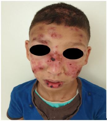
A 9-year-old previously healthy boy was admitted to our hospital with a 3-week history of eruption on the face, trunk and upper extremities associated with fever (38.5°C) and arthralgia.Physical examination revealed significant Centro facial oedema with confluent infiltrated erythematous plaques in a cocarde surmounted by crusts on the face (forehead, eyelid, cheeks, nose, and ear pinna) (Figure 1), trunk and palmar surface of both hands. Mucosal examination revealed multiple erosions over hard palate, hemorrhagic crust over lips and nasal mucosa (Figure 1), genital mucosa is intact. On the scalp, there are alopecic plaques with erythematous background of variable diameter. Systemic examination revealed no significant abnormalities. Investigation reports revealed anemia with hemoglobin ofvv 10.3 g/dL, lymphopenia 1000/ ml, neutropenia 1500 /mL and an erythrocyte sedimentation rate of 65 mm/h. A positive direct coombs test. The immunological tests showed a positive speckled ANA 1280, positive anti-DNA antibodies 80 and RF of 64 UI/mL, C4< 0, 06 g/L (0, 13–0, 57 g/L), C3 0, 38 g/L (0, 80–1, 90 g/L) and positive anti-Ro/SS-A. Kidney function was normal and proteinuria was negative; Epstein–Barr virus, cytomegalovirus, parvovirus B19, human immunodeficiency virus, and hepatitis B and C virus infections were ruled out. A skin biopsy was taken from one of the lesions and showed interface dermatitis with apoptotic keratinocytes, superficial and deep perivascular and periadnexal lymphoid infiltrate, with mucin deposition. Direct immunofluorescence was negative. Based on the clinical symptoms, laboratory tests, and histopathological examination, we made the diagnosis of Rowell’s syndrome. Therapy was started with prednisone 30 mg/day and hydroxychloroquine 200 mg/day with improvement.
Discussion
Rowell Syndrome (RS) is an unusual presentation of lupus erythematosus with erythema multiforme-like lesions associated with specific serological changes. Rare syndrome to date, there are 95 cases reported in the literature; of these, only seven cases are paediatric patients [4]. The association between LE and EM was described as a distinct feature by Neville Rowell et al. in 1963, including: Erythema multiforme lesions associated with discoid lupus erythematosus and specific immunological abnormalities (antinuclear antibodies in speckled fluorescence, serum antibodiesanti-La/SSB antibodies and the presence of a rheumatoid factor ) [2]. In 1995, Lee et al. proposed to introduce the presence of frostbite among the diagnostic criteria [5]. Over the years, Rowell Syndrome has gradually been described in association with all types of lupus [6]. In 2000, Zeitouni proposed precise diagnostic criteria for this entity, including three major criteria: presence of lupus erythematosus (systemic, subacute or discoid), erythema polymorphic lesions, presence of anti-nuclear speckled fluorescence antibodies, and three minor criteria: presence of anti-Ro/SSA or La/SSB antibodies, rheumatoid factor (RF) or frostbite [7]. Rowell syndrome is often preceded by a history of LE, but it has been reported to be its first sign [8]. It is important to exclude triggers for EM before con considering a diagnosis of RS. In this case, no triggers for EM were identified, and our patient met the major criteria of having systemic lupus erythematosus, erythema multiforme lesions, and the presence of speckled fluorescence antinuclear antibodies, as well as two other criteria, namely anti-Ro/SSA antibodies and a positive ( RF), we consider our case to be a classic RS. The histological findings of RS are controversial. For Torchia et al. There are no significant histological differences between Cutaneous Lupus Erythematosus (CLE) and EM lesions and the presence of necrotic keratinocytes is not specific to EM as it can also be found in CLE lesions [9]. Although diagnostic criteria have been established in the literature, the reality of this entity is still disputed [10]. The annular and targetoid appearance of the skin lesions can be confused with subacute lupus erythematosus and erythema multiforme. Some authors have suggested that lupus erythematosus with EM-like rashes designated as RS represent a subset of SCLE, rather than a separate entity. Others consider Rowell’s syndrome to be a singular entity [7,11]. Therapy schemes for Rowell’s disease include oral prednisone in medium and high doses, in combination with azathioprine or antimalarials such as chloroquine or hydroxychloroquine [13].
Declarations
Conflict of interest: None.
Funding source: None.
Consent statement: Written informed consent was obtained from both patients for publication of this case report and accompanying images.
References
- Shadid NH, Thissen CACB, van Marion AMW, Poblete-Gutiérrez P, Frank J, et al. Lupus erythematosus associated with erythema multiforme: Rowell’s syndrome. Int J Dermatol. 2007; 46: 30‑32.
- Rowell NR. Lupus Erythematosus and Erythema Multiforme-like Lesions: A Syndrome With Characteristic Immunological Abnormalities. Arch Dermatol. 1 août 1963; 88: 176.
- Shteyngarts AR, Warner MR, Camisa C. Lupus erythematosus associated with erythema multiforme: Does Rowell’s syndrome exist? Journal of the American Academy of Dermatology. mai 1999; 40: 773‑777.
- Aguirre-Martinez I, Vélez-Tirado N, García-Romero MT, Rodríguez-Lozano AL, Corcuera-Delgado CT, et al. Rowell syndrome complicated with macrophage activation syndrome in a child. Lupus. 2019; 28: 1716‑1721.
- Lee S, Schloss E, Kowichi J. Rowell’s syndrome: A case report with subacute cutaneous lupus erythematosus and erythema multiforme. Can J Dermatol. 1989; 21: 374-377.
- Aydogan K, Karadogan S, Balaban Adim S, Tunali S, et al. Lupus erythematosus associated with erythema multiforme: Report of two cases and review of the literature. J Eur Acad Dermatol Venerol. 2005; 19: 621‑627.
- Zeitouni NC, Funaro D, Cloutier RA, Gagné E, et al. Claveau J. Redefining Rowell’s syndrome: REDEFINING ROWELL’S SYNDROME. British Journal of Dermatology. févr. 2000; 142: 343‑346.
- Gallo L, Megna M, Festa B, Stellato P, di Pinto R, et al. Rowell Syndrome: A Diagnostic Challenge. J ClinAesthetDermatol. avr 2020; 13: 40‑42.
- Torchia D, Romanelli P, Kerdel FA. Erythema multiforme and Stevens-Johnson syndrome/toxic epidermal necrolysis associated with lupus erythematosus. Journal of the American Academy of Dermatology. 2012; 67: 417‑421.
- Schissler C, Banea S, Tortel MC, Mahé A. Un nouveau cas de syndrome de Rowell. Annales de Dermatologie et de Vénéréologie. avr 2017; 144: 263‑267.
- Massone, A. Parodi, A. Rebora C. Erythema Multiforme-likeSubacuteCutaneous Lupus Erythematosus: A New Variety? ActaDermato-Venereologica. 2000; 80: 308‑309.
- Shadid NH, Thissen CACB, van Marion AMW, Poblete-Gutiérrez P, Frank J, et al. Lupus erythematosus associated with erythema multiforme: Rowell’s syndrome. Int J Dermatol. 2007; 46: 30‑32.
- Bonciolini V, Antiga E, Caproni M, Fabbri P. Rowell syndrome: Does it exist? Clin Exp Dermatol. janv 2014; 39: 58‑58.