
Journal of Clinical Images and Medical Case Reports
ISSN 2766-7820
Case Report - Open Access, Volume 4
Onychodystrophy complicating bilateral linear
porokeratosis of mibelli: A rare association
Sokaina Chhiti*; Hanane Baybay; Fatima Zahrahashas; Zakia Douhi; Meriam Soughi; Sara Elloudi; Fatima Zahra Mernissi
Department of Dermatology CHU HASSAN II FES, Morocco.
*Corresponding Author : Chhiti Sokaina
Resident in the Dermatology Department, HASSAN II University Hospital, Fez, Morocco.
Phone: +21 2698308829;
Email: sokaina.chhiti@usmba.ac.ma
Received : Jan 23, 2023
Accepted : Mar 28, 2023
Published : Apr 04, 2023
Archived : www.jcimcr.org
Copyright : © Sokaina C (2023).
Abstract
Introduction: Porokeratosis is a specific disorder of keratinization whose diagnosis is based on clinical and histological findings. It presents in 5 clinical forms of which linear porokeratosis with nail dystrophy remains a rare entity.
Observation: We report the sporadic case of a 23-year-old girl who presented since childhood with linear porokeratosis lesions with nail dystrophy in an acral, bilateral and symmetrical pattern.
Discussion: Linear porokeratosis with nail dystrophy is a rare entity that can cause irreversible onychodystrophy and disabling underlying bone deformity.
Conclusion: The interest of early diagnosis and management to prevent and stop the evolution towards bone and nail destruction and deformation.
Keywords: Porokeratosis; Linear; Onychodystrophy; Cornoïd lamella; Bilateral; Acral.
Citation: Chhiti S, Baybay H, Zahrahashas F, Douhi Z, Soughi M, et al. Onychodystrophy complicating bilateral linear porokeratosis of mibelli: A rare association. J Clin Images Med Case Rep. 2023; 4(4): 2355.
Introduction
Porokeratosis is a specific hereditary disorder of keratinization that manifests itself clinically as well-defined annular or linear keratotic plaques of various sizes and shapes with the presence of cornoid lamellae on histology. There are 5 types of porokeratosis of which the linear form with nail dystrophy remains a rarely reported entity [1,2]. We report a rare case of linear porokeratosis affecting bilaterally and symmetrically both extremities with nail dystrophy in a 23-year-old girl.
Observation
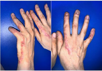
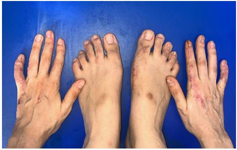
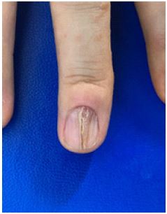
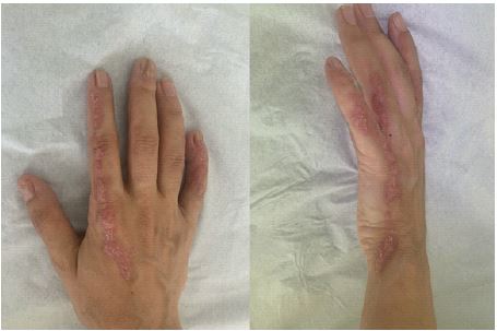
23-year-old girl, from a first-degree consanguineous marriage, without any notion of similar case in the family or trauma or neoplasia, who presents since childhood asymptomatic rough lesions on the hands and feet progressively increasing for which many traditional herbal treatments were applied without improvement. The dermatological examination showed multiple juxtaposed and confluent keratotic papules with irregular contours, crusty surface, erythematous and slightly atrophic border, linearly arranged along Blaschko’s lines on the anterior surfaces of the hands and feet reaching the lateral edges of the fingers (Figures 1,2) with onycholysis, onychorrhexis, hyperkeratosis and multiple longitudinal fissures on the nails of the 2 middle fingers, the little fingers and the thumb of the right hand (Figure 3).
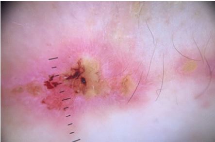
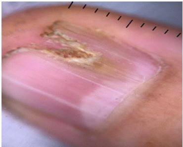
On dermoscopy: An erythematous background, spotty vascularity and crusts (Figure 4). At the nail level: onycholysis, paronychia and xantonychia (Figure 5). Histological examination confirmed the diagnosis of porokeratosis. X-ray of the hands was abnormal. The patient was initially put on oral isotretinoin at a dose of 20 mg/day for 2 months without improvement, then the patient benefited from 3 sessions of ablative fractional laser combined with salicylated vaseline with partial improvement (Figure 6).
Discussion
Porokeratosis is classically described by Mibelli [3] and Respighi [4] in 1893, as a disorder of keratinization characterized clinically by asymptomatic lesions made of centrifugal annular hyperkeratotic papules or plaques with an atrophic center and histologically by the presence of characteristic cornoïd lamellae. There are 5 clinical variants [1,2]. Some of them were noted destructive and others were superficial non-atrophic lesions [5]. Linear porokeratosis is a rare variant (3.5% to 16.7%) strictly unilateral and following Blaschko’s lines with identical Mibelli morphology [6]. This was not the case in our patient who presented with a bilateral linear acral arrangement without atrophic center or centrifugal evolution. Usually this type of porokeratosis is sporadic but sometimes it occurs as an autosomal dominant hereditary disease with frequent involvement of the extremitieusually appearing at birth or in childhood [7] like our patient.
Nail involvement in linear porokeratosis is extremely rare [7,8]. However, when they are present, they are mainly in linear form with fissures, longitudinal striae, anonymity and pterygium formation [7-9]. Although the exact pathogenesis of porokeratotic nail is still poorly elucidated, these changes can be explained by damage to the nail bed and/or nail matrix by abnormal clonal keratinocytes resulting in onychodystrophy. In a patient with generalized porokeratosis with oral lesions, the nail involvement resembled changes in onychomycosis [10] such as the case of our patient. Some authors have observed́ shortening, distal digital narrowing, bone resorption, flexion contracture, and mutilation with loss of the fingers involved above the linear porokeratosis lesions [7] fortunately our patient had no underlying destruction.
Linear porokeratosis lesions are more prone to malignant degeneration, especially in Bowen’s disease, squamous cell carcinoma and rarely in basal cell carcinoma. This can be explained by the loss of heterozygosity or by the occurrence of chronic scarring areas such as anonymity [11]. However, no such cases have been reported to date,
Despite the existence of several treatment options such as cryotherapy, topical 5FU, topical retinoids, lasers, shaving excision and dermabrasion, none of them has showń promising efficacý and long-term remission [9,12]. However, there is no clear consensus on treatment, especially when nail dystrophy is associated with porokeratosis [13]. Reported successful treatment with a combination of imiquimod and efudex. Although partial improvement was achieved with isotretinoin, we then had greater improvement with the use of an ablative laser combined with salicylated vaseline after 6 months.
Conclusion
In conclusion, we suggest that Porokeratosis involving the distal extremities, as in our case, may lead to onychodystrophy with underlying disabling bone involvement in some cases hence the need for recognition of this entity in order to avoid early destructive changes in the disease. In our patient, long-term follow-up will be necessary to detect malignant changes as early as possible.
Declarations
Consent: The examination of the patient was carried out in accordance with the principles of the Declaration of Helsinki.
Conflicts of interest: The authors declare no conflict of interest.
References
- Das A, Vasudevan B, Talwar A. Porokeratosis: An enigma beginning to unravel. Indian J Dermatol Venereol Leprol. 2022; 88: 291-299.
- Lorenz GE, Ritter SE. Linear porokeratosis: A case report and review of the literature. Cutis. 2008; 81: 479-483.
- Mibelli V. Contributo allo etudiodellaipercheratosi dei canalisudoriferi (porokeratosis). Giornale Italianodella Tuberculosiedellamalattie Thorace. 1893; 28: 313-355.
- Respighi E. Di unaipercheratosi non ancoradescritta. G Ital Mal Veneree Pelle. 1893; 28: 356-386.
- Gutierrez EL, Galarza C, Ramos W, et al. Facial porokeratosis: A series of six patients. Australas J Dermatol. 2010; 51: 191-194.
- Peyrière H, Dereure O, Breton H, et al. Variability in the clinical pattern of cutaneous side-effects of drugs with systemic symptoms: Does a DRESS syndrome really exist?. Br J Dermatol. 2006; 155: 422-428.
- Pawar M. Onychodystrophy due to porokeratosis of Mibelli: A rare association. Acta Dermatovenerol Alp PannonicaAdriat. 2017; 26: 51-52.
- Kohara Y, Takeo T, Oshima Y, Akita Y, Tamada Y, Watanabe D, et al. Linear porokerato- sis with nail dystrophy. Eur J Dermatol. 2011; 21: 625–626.
- Kohara Y, Takeo T, Oshima Y, et al. Linear porokeratosis with nail dystrophy. Eur J Dermatol. 2011; 21: 625-626.
- Kobayasi T. Generalized porokeratosis of Mibelli with lesions of buccal mucosa and of the nails. Jap J Derm Urol. 1934; 36: 439–442.
- Yendo TM, Gabbi TVB, Nico MMS. Porokeratosis of the Nail Unit: Case Series and Review. Skin Appendage Disord. 2021; 7: 489-492.
- Kanitakis J. Porokeratoses: An update of clinical, aetiopathogenic and therapeutic features. Eur J Dermatol. 2014; 24: 533-544.
- Vij A, Doherty SD, Leleux TM, Hsu S. Disseminated porokeratosis of Mibelli: A case report. Dermatology Online Journal. Dermatol Online J. 2010; 16: 12.