Journal of Clinical Images and Medical Case Reports
ISSN 2766-7820
Case Report - Open Access, Volume 4
Meningomyelocele - A classical clinical and radiological image
Sri Sita Naga Sai Priya K*; Meshram RJ2
1Junior Resident, Datta Meghe Institute of Higher Education and Research, Wardha, India.
2Associate Professor, Datta Meghe Institute of Higher Education and Research, Wardha, India.
*Corresponding Author : Sri Sita Naga Sai Priya K
Junior Resident, Datta Meghe Institute of Higher Education and Research, Wardha, India.
Email: saipriyakalakota0596@gmail.com
Received : Mar 25, 2023
Accepted : Apr 13, 2023
Published : Apr 20, 2023
Archived : www.jcimcr.org
Copyright : © Priya KSSNS (2023).
Keywords: Neural Tube Defects; Congenital malformation; Lumbosacral swelling.
Citation: Priya KSSNS, Meshram RJ. Meningomyelocele - A classical clinical and radiological image. J Clin Images Med Case Rep. 2023; 4(4): 2379.
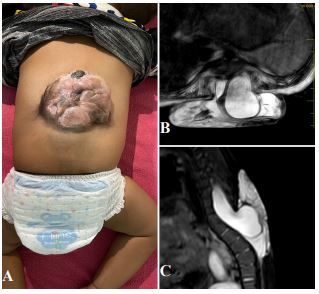
Clinical image description
Neural tube defects (NTD) are one of the most common congenital malformations, second only to congenital heart disease (CHD). The most common types of NTDs are anencephaly and spina bifida. NTDs can be open or closed. Unlike some other congenital defects, NTDs can usually be prevented. When women take the recommended amounts of folic acid (FA) during the periconceptional period and the first trimester, they can significantly lower the risk of NTDs. The incidence of NTDs is higher in India as compared to developed countries. NTDs are associated with various severe complications, like cognitive delay, seizures, paraparesis, neurogenic bladder, etc. The clinical image is of a 2-year-old male born with swelling over the mid-back and no movement or power in bilateral lower limbs (Figure 1A). The patient was brought in at the age of 2 years for further management. The patient had a gross developmental delay with macrocephaly. The MRI done was suggestive of a meningomyelocele seen at the levels of L5, S1, and S2 with herniation of the meninges and neural tissue/nerve roots (Figures 1B and 1C). The spinal cord was tethered and lying low (up to L4-5). A subsequent MRI of the brain was suggestive of aqueductal stenosis with ventricular dilatation with no other significant anomaly. Other systemic anomalies known to be associated were ruled out. NTDs are known as one of the few birth defects for which primary preventive strategies are available and effective. Periconceptional folate intake can prevent about 70% of NTDs. A dose of 400 mcg/day is routinely prescribed to all pregnant ladies, starting when the couple is ready to conceive and is continued throughout the first trimester. If a mother has a previous history of NTD in a child is given 10 times the dose (5 mg/day). This image gives a diagnosis of myelomeningocele with a differential diagnosis of meningocele, encephalocele, or spina bifida.