
Journal of Clinical Images and Medical Case Reports
ISSN 2766-7820
Clinical Image - Open Access, Volume 4
Rare case of femoral shaft fracture in osteopetrosis
Yassine Ben Bouzid*; Marouane Dinia; Rida-Allah Bassir; Monsef Boufettal; Jalal Mekkaoui; Mohamed Kharmaz; Moulay Omar Lamrani; Mohamed Saleh Berrada
Department of Orthopaedic and Trauma Surgery, Ibn Sina University Hospital, Rabat, Morocco.
*Corresponding Author : Yassine Ben Bouzid
Department of Orthopaedic and Trauma Surgery, Ibn Sina University Hospital, Rabat, Morocco.
Email: yassine.benbouzid2@gmail.com
Received : Mar 21, 2023
Accepted : Apr 18, 2023
Published : Apr 25, 2023
Archived : www.jcimcr.org
Copyright : © Bouzid BY (2023).
Citation: Bouzid BY, Dinia M, Bassir RA, Boufettal M, Mekkaoui J, et al. Rare case of femoral shaft fracture in osteopetrosis.J Clin Images Med Case Rep. 2023; 4(4): 2386.
Introduction
First described by Albert Schonberg in 1904 [1], osteopetrosis is a group of sclerosing bone dysplasias characterized by generalized skeletal densification due to decreased osteoclast mediated resorption [2].
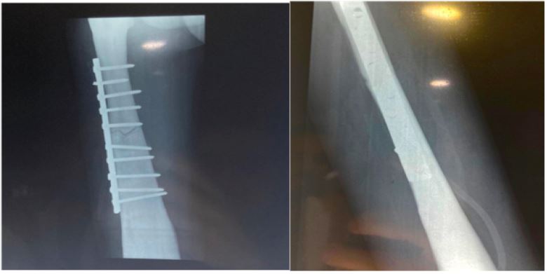
The management of fractures in this patient population is unique. Centromedullary nailing cannot be used in the vast majority of cases because of a narrow canal. Thus, Open Reduction And Internal Fixation (ORIF) remains the most suitable method in these patients [3].
Visual case discussion
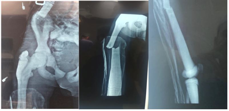
A 18 years old patient, suffering from osteopetrosis with cecity and bone marrow deficiency, was admitted to the emergency room of the Ibn Sina University Hospital in Rabat for a mid-shaft fracture of the right femur following a minor fall on the thigh.
The clinical examination revealed a patient with functional impotence of the right lower limb; a deformity of the thigh; the examination of the hip and knee was normal. The pulses were well perceived and there was no sensory-motor deficit.
On standard radiography, a femoral shaft fracture with medullary canal insufficiency and generalized bone density increase was observed.
The patient was admitted to the operating room where he underwent surgical management by ORIF. The postoperative follow-up was simple.
References
- Albers-Schonberg HE. Rontgenbilder einer seltenen Knockenerkrankung. Munch Med Wochenschr. 1904; 51: 365-368.
- Marks SC. Pathogenesis of osteopetrosis in the rat: Reduced bone resorption due to reduced osteoclast function. Am J Anat. 1973; 138: 165-178.
- Hao Yang , Guo Xi Shao , Zhen Wu Du , Zheng-Wei Li, et al. Treatment for subtrochanteric fracture and subsequent nonunion in an adult patient with osteopetrosis: A case report and review of the literature.