
Journal of Clinical Images and Medical Case Reports
ISSN 2766-7820
Short Report - Open Access, Volume 4
Diffuse osteosclerosis of axial and appendicular skeleton
is rare in renal osteodystrophy: Case report
*Corresponding Author : Amit Kumar
Department of Radio-diagnosis, IGIMS, Patna, India.
Email: amitragini99@gmail.com,
amitmd2008@gmail.com
Received : Mar 20, 2023
Accepted : Apr 19, 2023
Published : Apr 26, 2023
Archived : www.jcimcr.org
Copyright : © Kumar A (2023).
Abstract
Diffuse osteosclerosis of axial and appendicular skeleton is an unusual manifestation of renal osteodystrophy. Symmetrical involvement of multiple bones is the finding. The more common encountered feature is rugger jersey appearance of spine due to affected vertebral end plates.
We present a case with chronic kidney disease and renal osteodystrophy manifesting in the form of diffuse osteosclerosis.
Keywords: Chronic kidney disease; Renal osteodystrophy; Diffuse osteosclerosis; Axial and appendicular skeleton.
Citation: Kumar A. Diffuse osteosclerosis of axial and appendicular skeleton is rare in renal osteodystrophy: Case report. J Clin Images Med Case Rep. 2023; 4(4): 2387.
Introduction
Renal osteodystrophy is a term comprising all disorders of calcium and phosphate metabolism along with abnormalities of musculoskeletal system in a patient with chronic kidney disease [1]. It is an amalgam of pathological conditions comprising hyperparathyroidism and osteomalacia. The finding may involve both spectrum i.e. Osteopenia and osteosclerosis [2]. Also, sclerosis involving only the bone ends is an unusual finding and it must be differentiated from osteonecrosis [3].
This case demonstrated the unusual spectrum of renal osteodystrophy with diffuse osteosclerosis of multiple bones.
Case report
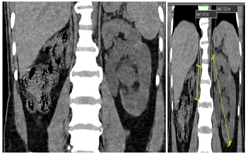
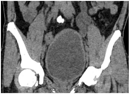
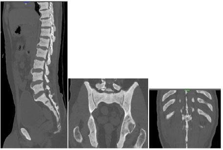
A 45-year-old male came to the Department of Medicine for regular follow up for impaired kidney function test. Routine blood investigations showed raised serum creatinine level. He was referred to the department of Radiodiagnosis for NCCT KUB study. It showed a small right kidney- likely atrophied (Figure 1). Left kidney showed compensatory enlargement with moderate hydronephrosis (Figure 1). The left ureter was also dilated in its whole extent (Figure 2). The bladder was distended with mildly thickened and irregular wall (Figure 2) with urinary bladder diverticula. Bone window showed marked, diffuse osteosclerosis of multiple vertebrae, ribs and hip bones (Figure 3).
Discussion
In a long-standing case of renal insufficiency, secondary hyperparathyroidism occurs due to parathyroid cell hyperplasia [4]. This increased secretion of parathormone is due to decreased level of serum calcium caused by increased serum phosphate levels and decreased 1.25(OH)2. Vitamin D as a consequence of impairment of renal parenchyma [5].
Osteosclerosis is a common manifestation of chronic renal insufficiency. However, it usually involves metaphysis of long bones or superior and inferior end plates of vertebrae, resulting in classical appearance of ‘rugger jersey vertebrae’ which is quite pathognomic of hyperparathyroidism, particularly due to chronic renal failure. But involvement of bone ends is quite unusual [6-9].
The main differentials of rugger jersey spine include Paget’s disease which is characterised by picture frame vertebra [8]. And Osteopetrosis, characterised by Sandwich vertebra [10].
Osteosclerosis predominantly involves spine followed by pelvis, ribs and skull. The accentuated radiodensity of bones is attributed to increased trabecular bone mass [3].
In some cases, haematological decompensation may occur due to compromised marrow function.
Bone biopsy is the gold standard to confirm the diagnosis of Renal osteodystrophy. Radiological investigations help in early diagnosis and aids in follow up of patients in monitoring the effects of therapy.
Conclusion
Renal osteodystrophy is commonly encountered in the patients with chronic kidney disease but they present as sclerosis of the end plates of the vertebral bodies. It is also important to know the other varied and unusual presentation in CKD. Here, we report a case with rare presentation of bone involvement in the form of diffuse osteosclerosis.
References
- Oprisiu R, Hottelart C, Ghitsu S, Said S, Westeel PF, Moriniere P, et al. Renal osteodystrophy (1): invasive and non-invasive diagnosis of its pathologic varieties. Nephrologie. 2000; 21: 229-237.
- Eastwood JB. Renal osteodystrophy--a radiological review. CRC Crit Rev Diagn Imaging. 1977; 9: 77-104.
- Lewis VL, Keats TE. Bone end sclerosis in renal osteodystrophy simulating osteonecrosis. Skeletal Radiol. 1982; 8: 275-278.
- McHenry CR, Wilhelm SM, Ricanati E. Refractory renal hyperparathyroidism: Clinical features and outcome of surgical therapy. Am Surgeon. 2001; 67: 310-317.
- Fournier A, Oprisiu R, Hottelart C, Yverneau PH, Ghazali A, et al. Renal osteodystrophy in dialysis patients: diagnosis and treatment. Artif Organs. 1998; 22: 530-557.
- Eastwood JB Renal osteodystrophy - A radiological review. CRC Crit Rev Diagn Imaging. 1977; 9: 77-104.
- Garver P, Resnick D, Niwayama G, Guerra J Jr. Epiphyseal sclerosis in renal osteodystrophy simulating osteonecrosis. AJR 1981; 136: 1239-1241.
- Kirkland JD, O’Brien WT. Osteopetrosis – Classic Imaging Findings in the Spine. Journal of Clinical and Diagnostic Research. J Clin Diagn Res. 2015; 9: TJ01- TJ02.
- Martell BS, Dyer RB The rugger jersey spine. Abdominal Imaging. 2015; 40: 3342-3343.
- Wittenberg A. The Rugger Jersey Spine Sign. Radiology. 2004; 230: 491-492.