
Journal of Clinical Images and Medical Case Reports
ISSN 2766-7820
Case Report - Open Access, Volume 4
Revolutionizing oral surgery: The benefits of diode
laser for soft tissue lesion removal
*Corresponding Author : Moamen M Sarhan
Assistant Lecturer of Prosthodontics, Faculty of Dentistry, Damanhour University (and PhD Student in Prosthodontic Department Alexandria University), Egypt.
Email: moamen.sarhan@yahoo.com
Received : Mar 20, 2023
Accepted : Apr 20, 2023
Published : Apr 27, 2023
Archived : www.jcimcr.org
Copyright : © Sarhan MM (2023).
Abstract
Epulis fissuratum is a condition where the tissues in the mouth become overgrown due to long-term irritation caused by poorly fitting dentures. The cause of this condition is ongoing irritation and damage to the tissue, which is most commonly seen in the areas surrounding the labial vestibule. In this report, a 57-year-old man had an excessive growth of soft tissue in the lower premolar region. The growth was removed using a diode laser, and the healing process was straightforward, with no need for stitches, periodontal packs, or pain relievers. Follow-up examinations showed no signs of regrowth.
Keywords: Denture induced hyperplasia; Diode laser; Epulis fissuratum.
Citation: Sarhan MM. Revolutionizing oral surgery: The benefits of diode laser for soft tissue lesion removal. J Clin Images Med Case Rep. 2023; 4(4): 2390.
Introduction
Epulis fissuratum is an oral pathologic condition that appears in the mouth as an over growth of fibrous connective tissue, also known as inflammatory fibrous hyperplasia, denture epulis, or denture-induced fibrous hyperplasia, is a painless proliferation of gingiva [1].
Epulis fissuratum appears as a single or multiple fold of tissue that grown in excess around the alveolar vestibule that is firm in texture and has a normal color. The development of this condition is due to prolonged irritation caused by ill-fitting prostheses situated near the flange of the denture. Epulis fissuratum usually presents as a raised lesion that is frequently sessile and has a smooth surface. The color of the lesion looks like normal mucosa. The dimensions of the affected tissue varies widely, since almost the entire length of tissue around a denture can be affected [2]. More commonly found in women. While it can occur in either in lower jaw or upper, it is more frequently found on the labial sulcus. This condition is also vulnerable to fungal superinfection [3].
Epulis fissuratum can be treated with topical antifungal agents, complete surgical exision, and creation of a new acrylic complete denture with proper borders. Even though minimizing trauma and irritation from the denture cause minor reduction in the dimension of the lesion, definitive treatment typically involves conservative excisional surgery. Recurrence of the lesion is unlikely if the sources of trauma and/or the patient’s behaviours are removed and suitable prosthetic repair is provided [4].
Three common methods are used to remove overgrown tissues: scalpel, electrocautery, and laser. Several lasers, including the carbon dioxide laser, neodymium-doped yttrium aluminium garnet (Nd:YAG) laser, and erbium, chromium: yttrium, scandium, gallium, garnet (Er, Cr: YSGG) laser, have been employed for this purpose in the past. While CO2 and Nd:YAG lasers are known for their ability to remove soft tissue, they can also cause significant thermal effects on the targeted tissues [5-7].
The report documents a case in which a diode laser was used to treat a patient with epulis fissuratum. This laser has been shown efficient in removing soft tissue lesions in the oral cavity and performing gum-related procedures. Advantages of diode laser treatment include reduced bleeding during surgery, which leads to a clearer surgical field and shorter operation time. Additionally, the surgical site is disinfected, and the healing process is faster with little to no scarring, minimal postoperative pain, and a lower cost compared to other types of lasers [8-11].
Case report
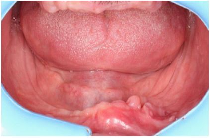
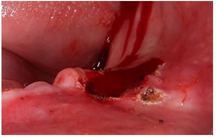
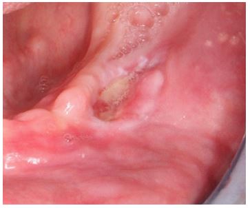
A 57-year-old man visited the Prosthodontics Department at Alexandria University, complaining of tissue overgrowth in the lower buccal vestibule that had been present for the past four months (Figure 1). Upon intraoral examination, a hyperplastic growth of soft tissue was observed extending from the canine to premolar on the buccal vestibule of the mandibular arch. The growth was related to the flange of a broken denture that did not fit properly.
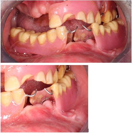
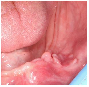
The growth appeared to be localized, pink, fibrous, and attached directly to the tissue without a stalk, with irregular borders present over the buccal vestibule of the mandibular arch. The patient reported having been using the broken denture for the past six months (Figures 2 and 3) and had no history of any relevant systemic disease.
Following a clinical examination, an evaluation of the patient’s history, and a review of the broken denture, a diagnosis of “epulis fissuratum” was made. The patient was scheduled for the removal of the lesion using a diode laser, and a new complete denture was to be created. The cause of the lesion was most likely the prolonged usage of an ill-fitting denture, resulting in chronic irritation of the tissues in the denture-bearing region. This resulted in a 5.8 mm by 15.4 mm region of soft tissue overgrowth (Figure 4). The patient was advised to remove the old denture and allow the affected tissue to rest for two weeks before undergoing an excisional removal of the lesion using a diode laser. To ensure safety, the region around the lesion was infiltrated with local anesthetic solution (Articaine Hydrochloride 68 mg/1.7 ml with Adrenaline 0.017 mg/1.7 ml), and the patient, assistant, and surgeon all wore laser safety glasses.
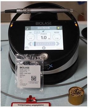
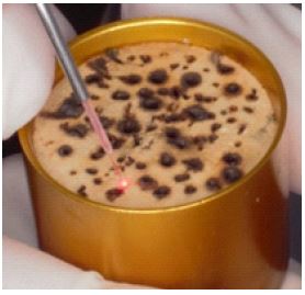
To remove the lesion, a diode laser (Epic x, Biolase, Inc. USA) with a wavelength of 940 nm and a laser classification of class IV was used in contact mode and continuous mode with an initiated tip (E4) at a power of 1 W, using a sweeping motion (Figures 5 and 6). All tissue tags were removed, and the surgical field was irrigated with normal saline. No sutures, periodontal pack, or stent were needed, and the wound was allowed to heal naturally (Figure 7). No drugs were given, and the patient was instructed to refrain from hot and spicy meals for three days. A follow-up assessment was conducted after one week, and the healing process was good and unremarkable (Figure 8).
Discussion
Inflammatory fibrous hyperplasia, also referred to as epulis fissuratum, is a medical condition in which there is an abnormal growth of fibrous connective tissue in the alveolar mucosa and vestibular area [1]. The most common cause of epulis fissuratumis associated with the edges of a denture that does not fit well. This can lead to increased keratinization and proliferation of connective tissues, which ultimately results in the development of epulisfissuratum [1].
Epulis fissuratum, a condition that can be treated through conventional excisional biopsy, electrosurgery, and laser techniques. The selection of treatment method depends on the size and extent of the lesion [5,6]. Laser therapy offers many benefits over other treatment options. It causes minimal bleeding, sterilizes the surgical site instantly, reduces bacteremia, and results in less trauma, swelling, and postoperative pain. In addition, laser therapy results in minimal wound contraction and scarring, which makes it an appealing choice for completely edentulous patients to avoid obliteration of the sulcus [9-11].
Conclusion
Diode laser is a highly effective and cost-efficient method for removing small soft tissue lesions in oral surgery. It is preferred for its ease of use, adequate coagulation, and ability to minimize swelling and pain, without the need for sutures. It is considered a safe, predictable, and superior alternative to conventional scalpel and other types of laser or electric scalpel due to its numerous advantages.
References
- Cawson RA, Odell EW. Essentials of oral pathology and oral medicine. Churchill Livingstone. 1998.
- Sagar K, Tandon S, Lamba AK, Yadav N. Diode laser assisted management of denture induced fibrous hyperplasia. Journal of Dental Lasers. 2013; 7: 77.
- Netto CH. Protese total imediata. 11th ed. Sao Paulo: Pancast Editorial. 1987; 107-134.
- Torres Domingo S, Bagán Sebastián JV, Jiménez Soriano Y, Poveda Roda R, Murillo Cortés J, et al. Benign tumors of the oral mucosa: A study of 300 patients.
- Eroglu CN, Tunc SK, Elasan S. Removal of epulisfissuratum by Er, Cr: YSGG laser in comparison with the conventional method. Photomedicine and laser surgery. 2015; 33: 533-539.
- Azma E, Safavi N. Diode laser application in soft tissue oral surgery. J Lasers Med Sci. 2013; 4: 206-211.
- Desiate A, Cantore S, Tullo D, Profeta G, Grassi FR, et al. 980 nm diode lasers in oral and facial practice: Current state of the science and art. Int J Med Sci. 2009; 6: 358-364.
- Amaral MB, de Ávila JM, Abreu MH, Mesquita RA, et al. Diode laser surgery versus scalpel surgery in the treatment of fibrous hyperplasia: a randomized clinical trial. Int J Oral Maxillofac Surg. 2015; 44: 1383-1389.
- Gargari M, Autili N, Petrone A, Ceruso FM, et al. Using laser diodes for the removal of a lesion of the oral mucosa. Case report. Oral Implantol (Rome). 2011; 4: 10-13.
- Cercadillo-Ibarguren I, España-Tost A, Arnabat-Domínguez J, Valmaseda-Castellón E, Berini-Aytés L, et al. Histologic evaluation of thermal damage produced on soft tissues by CO2, Er,Cr: YSGG and diode lasers. Med Oral Patol Oral Cir Bucal. 2010; 15: e912-e918.
- Chandna S, Kedige SD. Evaluation of pain on use of electrosurgery and diode lasers in the management of gingival hyperpigmentation: A comparative study. J Indian Soc Periodontol. 2015; 19: 49-55.