Journal of Clinical Images and Medical Case Reports
ISSN 2766-7820
Clinical Image - Open Access, Volume 4
Eye shaped sub-hyaloidal hemorrhage. What does
the eye reveal about the body?
Kawtar Bouirig*; Lalla Ouafacherkaoui
Ophthalmology Department “A”, Ibn Sina University Hospital (Hôpital des Spécialités), Mohammed V University, Rabat, Morocco.
*Corresponding Author : Kawtar Bouirig
Ophthalmology Department “A”, Ibn Sina University Hospital (Hôpital des Spécialités), Mohammed V University, Rabat, Morocco.
Email: bouirigkawtar@gmail.com
Received : Apr 05, 2023
Accepted : Apr 21, 2023
Published : Apr 28, 20230
Archived : www.jcimcr.org
Copyright : © Bouirig K (2023).
Keywords: Retinal macroanevrysm; Sub-hyaloidal hemorrhage; Laser yag; Blood pressure.
Citation: Bouirig K, Ouafacherkaoui L. Eye shaped sub-hyaloidal hemorrhage. What does the eye reveal about the body? J Clin Images Med Case Rep. 2023; 4(4): 2392.
Description
A 62 year old female patient, with no previous history, consulted in emergency department for a sudden decrease in visual acuity of the right eye with a feeling of uneasiness and vertigo.
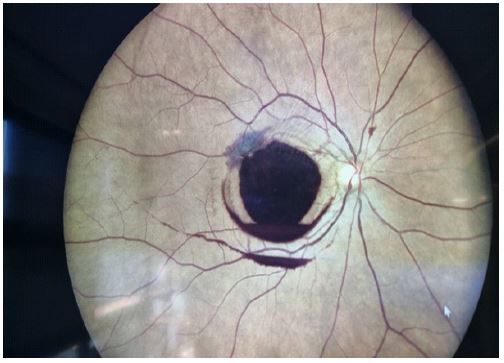
The Best Corrected Visual Acuity (BCVA) was measured at 10/10 according to the Snellen scale in the left eye and “counting fingers” in the right eye. The fundus examination showed a premacular, sub-hyloidal hemorrhage, taking the shape of an eye with a double crown developed at the expense of a superior temporal macroaneurysm.
Spectral-Domain Optical Coherence Tomography (SD-OCT) confirmed the premacular location of the blood, bordered in the superior temporal by an intraretinal hemorrhage containing an aspect of ruptured Retinal Arterial Acroaneurysm (RAAM) causing the sub-hyaloidal hemorrhage. The blood pressure was measured immediately, showing a high blood pressure of 170/80 mmhg. The patient was referred to his attending cardiologist for management.
As the visual acuity did not improve after 7 days, a hyaloidotomy with laser Yag was programmed and the visual acuity was restored to 7/10 one week after the laser.
Retinal Arterial Macroaneurysm (RAAM) is an acquired vascular pathology. Typically it is solitary, round or fusiform in shape, located on the path of the 4 main retinal arterial branches in the para-macular or posterior equatorial zone. The main risk factors are arterial hypertension and atherosclerosis. It is usually described in subjects over 60 years of age with a female predominance. Fibrinolytic disorders, Valsalva maneuvers, a drug induced origin should be considered as a differential diagnosis when searching for the etiology of a sub-hyaloid hemorrhage.