
Journal of Clinical Images and Medical Case Reports
ISSN 2766-7820
Clinical Image - Open Access, Volume 4
From inflammation to prolapse: Chronic cervicitis and the development of uterocervical prolapse- A clinical insight
Shweta Patel; Satish Choudhury*; Ajay Halder; Bharti Singh
Department of Obstetrics & Gynaecology, All India Institute of Medical Sciences, Bhopal, India.
*Corresponding Author : Satish Choudhury
Department of Obstetrics & Gynaecology, All India Institute of Medical Sciences, Bhopal, India.
Email: satishchoudhury9@gmail.com
Received : Jun 29, 2023
Accepted : Jul 12, 2023
Published : Jul 19, 2023
Archived : www.jcimcr.org
Copyright : © Choudhury S (2023).
Keywords: Chronic cervicitis; Uterovaginal prolapse.
Citation: Patel S, Choudhury S, Halder A, Singh B. From inflammation to prolapse: Chronic cervicitis and the development of uterocervical prolapse- A clinical insight. J Clin Images Med Case Rep. 2023; 4(7): 2505.
Description
Uterine prolapse is the herniation of the uterus into or beyond the vagina as a result of the failure of ligamentous and fascial supports. It is almost always accurately diagnosed by clinical examination but maybe sometimes confused with its differential diagnoses. One of the differential diagnoses of uterine prolapse is a cervical mass protruding through the introitus. At birth, the cervix and uterine body are of equal size; in adult women, the body measures two to three times the size of the cervix. In uterine prolapse, this ratio of the size of the uterine body to the cervix is not altered. We report a rare case of III-degree utero vaginal prolapse with a bulky cervix which could mimic a cervical mass protruding through the introitus.
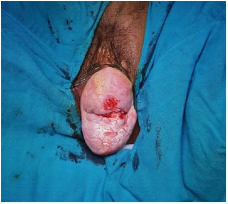
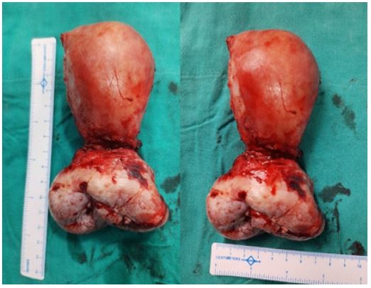
A 60-year female presented with a complaint of something coming out of her vagina for 2 years. She had no associated urinary or bowel complaints. On clinical examination, a pinkish-white structure of size 7.2 x 5.1 cm with a groove at its center was seen protruding through the introitus (Figure 1). Pelvic examination revealed this protruded structure to be a bulky cervix and the groove at its center being the external os. She was diagnosed with III-degree utero vaginal prolapse with a bulky cervix based on clinical examination findings. A vaginal hysterectomy was performed. On the measurement of the surgical specimen, the uterine body measured 7.0 x 4.0 cm and the size of the cervix was 7.2 x 5.1 cm (Figure 2). Histopathological examination revealed the findings consistent with normal uterus with chronic cervicitis.
Conclusion
Gynecologists should be aware of different clinical conditions which may mimic uterine prolapse. Uterine prolapse may be misdiagnosed as a cervical mass protruding through the introitus when the cervix is bulky and broad due to chronic cervicitis. A thorough clinical examination can accurately differentiate these two conditions and help in planning further management.
Conflicts of interest: None