
Journal of Clinical Images and Medical Case Reports
ISSN 2766-7820
Clinical Image - Open Access, Volume 4
Bullous leukemia cutis: A rare manifestation of acute myeloid leukemia
Vishnu Sharma1; Vansh Bagrodia2*; Tanishk Parchwani2
1Department of Clinical Hematology, SMS Medical College, Jaipur, India.
2Final year MBBS student, SMS Medical College, Jaipur, India.
*Corresponding Author : Vansh Bagrodia
Final year MBBS student, SMS Medical College,
Jaipur, India.
Email: vanshbagrodia@gmail.com
Received : Nov 08, 2023
Accepted : Dec 07, 2023
Published : Dec 14, 2023
Archived : www.jcimcr.org
Copyright : © Bagrodia V (2023).
Citation: Sharma V, Bagrodia V, Parchwani T. Bullous leukemia cutis: A rare manifestation of acute myeloid leukemia. J Clin Images Med Case Rep. 2023; 4(12): 2735.
Description
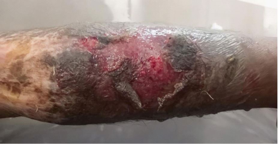
A 28-year-old male patient presented to our specialized unit with painful non-healing ulcers on both legs. The ulcers covered the lower 1/3rd part of the left leg and the upper 2/3rd part of the right leg (Figure 1). Blood counts showed hyperleukocytosis (87000/mm3) and smear showed 60% blasts. Imunophenotyping by flow cytometry suggested Acute Myeloid Leukemia.
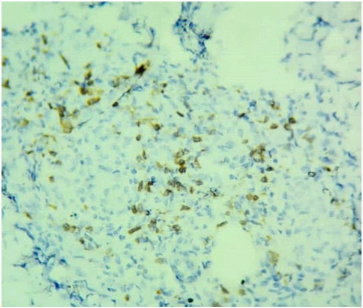
Punch biopsies of the skin lesions on both legs showed characteristic features of bullous leukemia cutis, including spongiosis, subcorneal bullae in the epidermis, necrosis and neutrophilic infiltration in the upper dermis, and infiltration by atypical leukemic cells in the deep dermis and subcutis positive for CD34 on immunohistochemistry (Figure 3). These findings confirmed the clinical diagnosis of myeloid sarcoma.
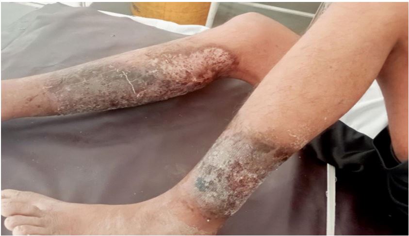
The patient underwent 7 + 3 (cytarabine + daunorubicin) therapy for AML and received supportive treatment for the ulcers, including analgesics, antibiotics, and antifungals. Due to the diagnosis of myeloid sarcoma, skin grafting was not recommended. The patient responded well to treatment, achieving complete response and bone marrow minimal residual disease (MRD) negativity (<0.1) (on flow cytometry) after completion of induction chemotherapy. The ulcers on both legs also showed marked improvement (Figure 2).
Bullous leukemia cutis represents a rare and challenging variant of leukemia cutis/myeloid sarcoma [1].
References
- Sandre M, Osmond A, Ghazarian D, Ghiasi N. Bullous leukemia cutis: A rare clinical subtype. Dermatology Online Journal. 2019; 25.