Journal of Clinical Images and Medical Case Reports
ISSN 2766-7820
Clinical Image - Open Access, Volume 5
A rare case of ethmoidal encephalocele with corpus callosal dysgenesis
Nayan Kabra1*; Atish Komwad2; Balaji Kombade3
1Junior Resident, VDGMC, Latur, India.
2Associate Professor, VDGMC, Latur, India.
3Professor and HOD, VDGMC, Latur, India.
*Corresponding Author : Nayan Kabra
Junior Resident, VDGMC, Latur, India.
Email: nankabra26@gmail.com
Received : Jun 10, 2024
Accepted : Jun 25, 2024
Published : Jul 02, 2024
Archived : www.jcimcr.org
Copyright : © Kabra N (2024).
Citation: Kabra N, Komwad A, Kombade B. A rare case of ethmoidal encephalocele with corpus callosal dysgenesis. J Clin Images Med Case Rep. 2024; 5(7): 3148.
Description
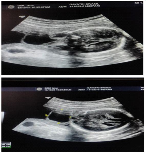
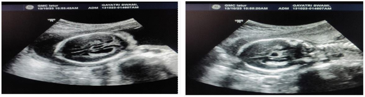
A 6 mm defect is seen over anterior aspect of face in between both orbits with a 2.6 x 2.5 cm sized cystic collection herniating through the defect possibly arising from ethmoid s/o Ethmoidal encephalocele. CSP (Cavum Septum Pellucidum) is not well visualised with bilateral parallel orientation of lateral ventricles. Third ventricle appears dilated. Splenium of corpus callosum not well visualised s/o Dysgenesis of corpus callosum. Cerebral hemispheres and Posterior fossa structure appears normal. A 6 mm defect is seen over anterior aspect of face in between both orbits with a 2.6 x 2.5 cm sized cystic collection herniating through the defect possibly arising from ethmoid s/o Ethmoidal encephalocele.