Journal of Clinical Images and Medical Case Reports
ISSN 2766-7820
Case Report - Open Access, Volume 2
Wandering spleen
Assaf Potruch*; Arnon Haran
Department of Medicine, Hebrew University-Hadassah Medical Center, Jerusalem, Israel.
*Corresponding Author: Assaf Potruch
Department of Medicine, Hebrew University-Hadassah
Medical Center, Jerusalem, Israel.
Email: poassaf@gmail.com
Received : Oct 25, 2021
Accepted : Dec 07, 2021
Published : Dec 14, 2021
Archived : www.jcimcr.org
Copyright : © Potruch A (2021).
Citation: Potruch A, Haran A. Wandering spleen. J Clin Images Med Case Rep. 2021; 2(6): 1479.
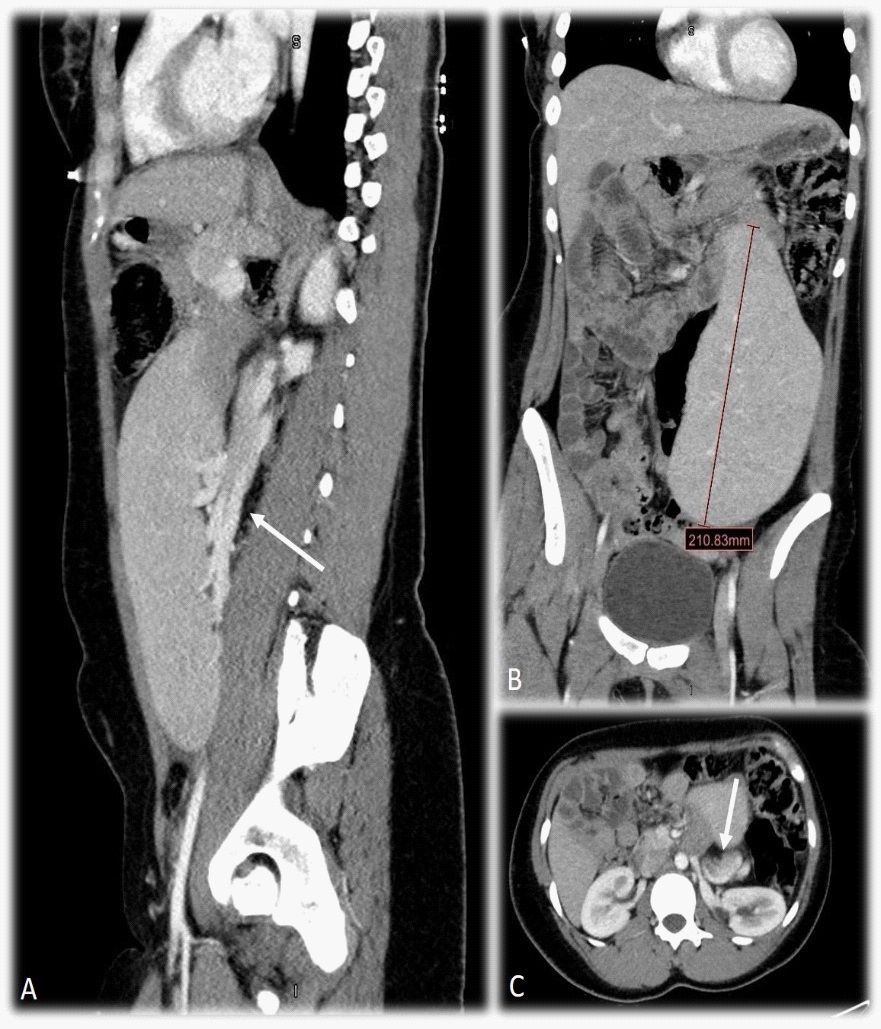
Clinical image description
A 25-year-old female presented to our hospital with severe abdominal pain of two days’ duration. A routine obstetric ultrasound several years prior had revealed an enlarged spleen, but she was otherwise healthy and had carried two uneventful pregnancies to term. Physical exam on arrival in the emergency room was notable for a tender mass in the subumbilical region. A CT scan revealed an enlarged and ectopic spleen (A, B) with twisting of its vascular pedicle and numerous venous collaterals adjacent to the splenic vein (A, C – arrows). The patient was admitted to the general surgery department but declined any surgical intervention following symptomatic improvement and was discharged. A so-called wandering spleen is a rare condition thought to be due to laxity of the splenic ligaments. Our patient’s acute presentation was likely due to a self-resolving episode of splenic vessel torsion, with previous episodes likely resulting in congestive splenomegaly