
Journal of Clinical Images and Medical Case Reports
ISSN 2766-7820
Clinical Image - Open Access, Volume 3
Beautiful aspect of hydatid cyst
Rita Oze Koudouhonon1*; Suzanne Rita Aubin Igombe1; Khadija Ben Elhosni2; Ittimade Nassar3; Nabil Moatassim Billal3
1Resident in Radiology, CHU Ibn Sina, Rabat, Morocco.
2Radiology Specialist, CHU Ibn Sina, Rabat, Morocco.
3Associate Professor in Diagnostic and Interventional Imaging, Central Radiology Department, CHU Ibn Sina Rabat, Morocco.
*Corresponding Author: Rita Oze Koudouhonon
Resident in Radiology, CHU Ibn Sina, Rabat,
Morocco.
Tel: +212 635349572;
Email: ritaoze23@gmail.com
Received : Dec 26, 2021
Accepted : Feb 09, 2022
Published : Feb 16, 2022
Archived : www.jcimcr.org
Copyright : © Koudouhonon RO (2022).
Keywords: hydatid cyst liver; CT; calcified floating membranes.
Citation: Koudouhonon RO, Igombe SRA, Elhosni KB, Nassar I, Billal NM, et al. Beautiful aspect of hydatid cyst. J Clin Images Med Case Rep. 2022; 3(1): 1663.
Description
Hydatid Disease (HD) is a mild endemic disease caused by ecchinococus granulosus. It can affect any system of the body, with hepatic predilection [1,2]. Most often asymptomatic, the literature describes 5 evolutionary stages ranging from simple cysts, with it poses a problem of differential diagnosis in its simple form to complicated forms that may or may not be calcified.
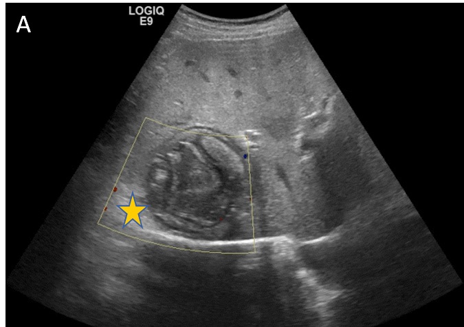
It is made up of three sofas; the pericyst, ectocyst, and endocyst. The CT scan plays an important role in imaging complicated hydatid cysts. The most common are rupture and superinfection. Occurring in 50 to 90% of cases, the rupture can be communicative or contained. When contained, the endocyst detaches from the pericyst appearing as a curvilinear structure or floating membranes which may subsequently calcify. This aspect is found on ultrasound, or on a CT scan [1,3]. The case on this hepatic ultrasound (Figure 1A) generated a grossly oval lesion, containing calcified serpiginous structures (floating membranes), generating shadow cones which mask the posterior reinforcement of this non-vascularized fluid mass on color Doppler (yellow star).
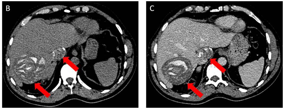
A complementary abdominal CT scan (Figure 2) in axial slices not evaluated (B), infectious (C), shows two hepatic lesions (segments 7 and 1) liquid, rounded with regular contours, containing calcified floating membranes (orange arrow).
References
- Alex Studemeister, Marcos N. Alvarez, Lucy Studemeister. Un patient atteint d’hémoptysie et le signe de la Camalote; Open Forum Infectious Diseases Forum ouvert Infect Dis. 2018; 5: dey286. Publié en ligne le 2 novembre 2018.
- Siham Eddeghai, Imane Eddoukani, Azzedine Diffaa, Khadija Krati. Kyste hydatique du foie: A propos d’un mode de révélation exceptionnel, Pan African Medical Journal. 2014; 18: 158.
- Mehta P, Prakash M, Khandelwal N. Manifestations radiologiques de la maladie hydatique et ses complications. Trop Parasitol. 2016; 6: 103-112.